The thought of an interview can be nerve-wracking, but the right preparation can make all the difference. Explore this comprehensive guide to Strong Understanding of Human and Animal Anatomy interview questions and gain the confidence you need to showcase your abilities and secure the role.
Questions Asked in Strong Understanding of Human and Animal Anatomy Interview
Q 1. Describe the layers of the skin in humans.
Human skin is a complex organ composed of three main layers: the epidermis, dermis, and hypodermis. Think of it like a three-layered cake, each layer with its own unique ingredients and function.
- Epidermis: This is the outermost layer, our body’s first line of defense. It’s thin but tough, made of stratified squamous epithelium. Its main role is protection against pathogens, dehydration, and UV radiation. The epidermis contains keratinocytes, which produce keratin, a tough protein that waterproofs the skin; melanocytes, which produce melanin, the pigment responsible for skin color and protection from UV damage; and Langerhans cells, which are immune cells.
- Dermis: This is the thick, middle layer, providing structural support and elasticity to the skin. It’s a dense connective tissue containing collagen and elastin fibers, giving it strength and flexibility. You’ll also find blood vessels, nerves, hair follicles, sweat glands, and sebaceous (oil) glands here. Imagine this layer as the skin’s plumbing and infrastructure system.
- Hypodermis (Subcutaneous Layer): This is the deepest layer, primarily composed of adipose (fat) tissue. It acts as insulation, cushioning, and energy storage. It also helps to anchor the skin to underlying muscles and bones. Think of this as the supportive base of the cake, providing a soft, cushioned foundation.
Understanding these layers is crucial for diagnosing skin conditions and developing effective treatments. For example, a sunburn affects primarily the epidermis, while deep wounds may penetrate through all three layers.
Q 2. Explain the function of the cardiovascular system in mammals.
The cardiovascular system in mammals is a closed circulatory system responsible for transporting blood, oxygen, nutrients, hormones, and waste products throughout the body. It’s like a sophisticated highway system, constantly delivering essential supplies and removing waste.
Its primary functions include:
- Oxygen transport: Blood carries oxygen from the lungs to the body’s tissues and cells.
- Nutrient delivery: Nutrients absorbed from the digestive system are transported to cells via the bloodstream.
- Waste removal: Metabolic waste products, such as carbon dioxide and urea, are transported to the lungs and kidneys for excretion.
- Hormone distribution: Hormones produced by endocrine glands are transported to their target organs via the blood.
- Temperature regulation: Blood helps to distribute heat throughout the body, maintaining a stable internal temperature.
- Immune defense: White blood cells, part of the immune system, are transported through the bloodstream to fight infections.
The heart acts as the central pump, propelling blood through arteries, capillaries, and veins. Dysfunction in this system can lead to various health problems, highlighting its importance.
Q 3. What are the main differences between the skeletal systems of birds and mammals?
The skeletal systems of birds and mammals, while both providing support and protection, differ significantly due to the unique adaptations of each group. Birds, designed for flight, have a skeletal system optimized for lightness and strength, while mammals show diversity depending on their locomotion and lifestyle.
- Weight Reduction: Bird bones are often pneumatic (hollow and filled with air sacs), significantly reducing their weight without compromising strength. Mammalian bones, while sometimes containing marrow cavities, are generally denser and heavier.
- Fusion of Bones: Birds have fused bones in several areas, such as the clavicles (forming the furcula or wishbone) and the bones of the pelvis and tail, providing rigidity and stability for flight. Mammalian skeletons tend to be more flexible.
- Keeled Sternum: Most birds possess a keeled sternum (breastbone), a large, prominent structure to which powerful flight muscles attach. Mammals lack this structure.
- Number of Bones: Birds generally have fewer bones than mammals of comparable size due to fusion and reduction.
These differences highlight the evolutionary adaptations driven by the need for flight in birds versus the varied terrestrial and arboreal lifestyles of mammals. Understanding these differences is crucial in comparative anatomy and paleontology, aiding in the reconstruction of extinct species and understanding evolutionary relationships.
Q 4. Illustrate the pathway of blood flow through the heart.
The pathway of blood flow through the heart is a continuous loop, ensuring oxygen-poor blood is oxygenated and oxygen-rich blood is delivered to the body’s tissues. Let’s follow the journey:
- Deoxygenated blood enters the right atrium from the body via the superior and inferior vena cava.
- The right atrium contracts, pushing blood through the tricuspid valve into the right ventricle.
- The right ventricle contracts, forcing blood through the pulmonary valve into the pulmonary artery.
- The pulmonary artery carries deoxygenated blood to the lungs, where it picks up oxygen and releases carbon dioxide.
- Oxygenated blood returns to the left atrium via the pulmonary veins.
- The left atrium contracts, pushing blood through the mitral valve into the left ventricle.
- The left ventricle contracts powerfully, sending oxygenated blood through the aortic valve into the aorta.
- The aorta distributes oxygenated blood throughout the body.
This cycle repeats continuously, maintaining the body’s oxygen supply. Any disruption to this precise pathway can lead to serious cardiovascular problems.
Q 5. Compare and contrast the digestive systems of herbivores and carnivores.
Herbivores and carnivores have digestive systems uniquely adapted to their diets. Herbivores, consuming plant matter, require systems to break down cellulose, while carnivores, eating meat, need systems to efficiently digest protein.
- Herbivores: Typically possess longer digestive tracts with specialized compartments like the rumen (in ruminants) or cecum (in hindgut fermenters). These chambers house symbiotic microorganisms that ferment cellulose, breaking it down into digestible nutrients. They often have larger, more complex stomachs and intestines to process the tough plant material.
- Carnivores: Generally have shorter, simpler digestive tracts. Their stomachs produce strong acids to break down proteins, and their intestines are relatively short because meat is easier to digest than plant matter. They rely on enzymes and efficient absorption processes.
For example, cows (herbivores) have a four-chambered stomach for cellulose digestion, while lions (carnivores) have a simple stomach with strong acids for protein digestion. These differences reflect the evolutionary adaptations to their specific diets.
Q 6. Explain the role of the nervous system in controlling movement.
The nervous system plays a crucial role in controlling movement, acting as the body’s command center. It’s a complex network of neurons that process information and coordinate actions.
The process generally involves:
- Sensory input: Sensory receptors detect stimuli (e.g., sight of an object you want to reach for). This information is transmitted to the central nervous system (brain and spinal cord).
- Integration: The brain processes this sensory information and formulates a motor plan (how to execute the movement).
- Motor output: The brain sends signals via motor neurons to muscles, instructing them to contract or relax.
- Muscle action: Muscles execute the motor plan, leading to the desired movement.
Different parts of the nervous system are involved, including the cerebellum (coordinating movement), the basal ganglia (initiating and controlling movement), and the motor cortex (planning and executing voluntary movements). Damage to any part of this pathway can result in impaired movement, highlighting its intricate and critical role.
Q 7. Describe the structure and function of a nephron.
The nephron is the functional unit of the kidney, responsible for filtering blood and producing urine. Imagine it as a tiny, highly efficient filtration plant within the kidney.
Its structure includes:
- Renal corpuscle: This is the filtering unit, composed of the glomerulus (a capillary network) and Bowman’s capsule (a cup-like structure surrounding the glomerulus). Blood pressure forces water and small solutes from the glomerulus into Bowman’s capsule.
- Renal tubule: This long, convoluted tube has three main sections: the proximal convoluted tubule (PCT), the loop of Henle, and the distal convoluted tubule (DCT). As filtrate flows through these sections, essential substances like water, glucose, and amino acids are reabsorbed back into the bloodstream. Waste products remain in the filtrate.
- Collecting duct: Several nephrons share a collecting duct, which further concentrates the urine by reabsorbing water. This concentrates the urine and regulates water balance.
The nephron’s function is to filter blood, reabsorb essential substances, and excrete waste products in the form of urine. This process is vital for maintaining the body’s fluid and electrolyte balance, regulating blood pressure, and removing metabolic waste. Kidney diseases often involve nephron dysfunction, illustrating their critical role in overall health.
Q 8. What are the key differences between human and animal respiratory systems?
While both human and animal respiratory systems share the fundamental goal of gas exchange (taking in oxygen and releasing carbon dioxide), significant differences exist based on species and lifestyle. Humans, as obligate terrestrial mammals, possess a highly developed system with a complex branching pattern of airways, maximizing surface area for efficient gas exchange in a stable atmospheric environment.
- Lung Structure: Human lungs are highly compartmentalized with alveoli, tiny air sacs, offering a vast surface area. Many animals, such as birds, have a more efficient system involving air sacs and unidirectional airflow, improving oxygen uptake during flight. Reptiles, amphibians, and some fish employ simpler lungs or cutaneous respiration (breathing through skin).
- Respiratory Rate & Control: Human respiratory rate is regulated by the brainstem, influenced by factors like oxygen and carbon dioxide levels. Animals may exhibit different respiratory patterns, influenced by factors such as body temperature, activity level, and even environmental oxygen availability.
- Respiratory Pigments: Humans utilize hemoglobin to carry oxygen in red blood cells. Animals might employ different respiratory pigments, like hemocyanin (in crustaceans and mollusks) or chlorocruorin (in some annelids), which bind and transport oxygen differently.
For example, a whale’s respiratory system is adapted for long dives, allowing for extended periods underwater by maximizing oxygen storage in their blood and muscles. In contrast, a hummingbird’s respiratory system is designed for the high metabolic demands of its rapid flight, characterized by an elevated respiratory rate and efficient oxygen extraction.
Q 9. Discuss the process of bone remodeling.
Bone remodeling is a continuous process of bone tissue renewal, involving the coordinated actions of two cell types: osteoclasts and osteoblasts. It’s essential for maintaining bone strength, calcium homeostasis, and adapting to mechanical stress.
- Osteoclasts: These large, multinucleated cells are responsible for bone resorption – the breaking down of old or damaged bone tissue. They secrete acids and enzymes that dissolve the bone mineral and matrix.
- Osteoblasts: These cells build new bone tissue by synthesizing and depositing new bone matrix, which eventually mineralizes. They also play a role in regulating the activity of osteoclasts.
The process is tightly regulated by hormones like parathyroid hormone (PTH), which stimulates bone resorption when calcium levels are low, and calcitonin, which inhibits it when calcium levels are high. Mechanical stress also influences remodeling; areas subjected to greater stress tend to have increased bone formation.
Imagine bone remodeling as a continuous cycle of demolition and construction. Old, weakened parts of the bone are broken down by osteoclasts, while osteoblasts rebuild it stronger and denser, leading to an overall healthier and more resilient skeleton.
Q 10. Explain the function of the lymphatic system.
The lymphatic system plays a crucial role in maintaining fluid balance, immunity, and lipid absorption. It’s a network of vessels, nodes, and organs that work together to transport lymph, a clear fluid containing white blood cells and other immune components.
- Fluid Balance: The lymphatic system collects excess interstitial fluid (fluid surrounding cells) that doesn’t re-enter the blood capillaries. This fluid is then filtered and returned to the bloodstream, preventing fluid buildup in tissues.
- Immunity: Lymph nodes act as filters, trapping bacteria, viruses, and other foreign substances. Lymphocytes (white blood cells) within the lymph nodes attack and destroy these invaders.
- Lipid Absorption: Lacteals, specialized lymphatic capillaries in the small intestine, absorb fats and fat-soluble vitamins that are not directly absorbed into the bloodstream.
A simple analogy: imagine the lymphatic system as a drainage system for your body, removing waste and harmful substances while also playing a vital role in your immune defense. Lymphedema, a condition caused by impaired lymphatic drainage, leading to swelling, highlights the crucial role of the lymphatic system in maintaining fluid balance.
Q 11. Describe the different types of muscle tissue and their functions.
Three main types of muscle tissue exist in the human body, each with distinct structural and functional properties:
- Skeletal Muscle: This is voluntary muscle, meaning its contraction is consciously controlled. It’s attached to bones via tendons, enabling movement. Skeletal muscle cells are long, cylindrical, and striated (having a striped appearance due to the arrangement of contractile proteins).
- Smooth Muscle: This is involuntary muscle, meaning its contraction is not consciously controlled. It’s found in the walls of internal organs like the stomach, intestines, and blood vessels. Smooth muscle cells are spindle-shaped and lack striations.
- Cardiac Muscle: This is involuntary muscle found only in the heart. It’s responsible for pumping blood throughout the body. Cardiac muscle cells are branched and striated, and they are interconnected by intercalated discs, specialized junctions that allow for coordinated contraction.
For instance, running involves the coordinated contraction of numerous skeletal muscles. Digestion relies on the rhythmic contractions of smooth muscles in the digestive tract. And a healthy heartbeat is maintained by the continuous, synchronized contractions of cardiac muscle.
Q 12. What are the major bones of the human skull?
The human skull is composed of numerous bones, broadly categorized into the cranium (protecting the brain) and the facial bones. Some major bones include:
- Frontal Bone: Forms the forehead and part of the eye sockets.
- Parietal Bones (2): Form the sides and roof of the cranium.
- Temporal Bones (2): Located on the sides of the skull, containing structures for hearing and balance.
- Occipital Bone: Forms the back of the skull and contains the foramen magnum (the opening where the spinal cord exits).
- Sphenoid Bone: A complex, butterfly-shaped bone situated at the base of the skull.
- Ethmoid Bone: Forms part of the nasal cavity and orbits.
- Maxilla (2): The upper jaw bones.
- Mandible: The lower jaw bone, the only movable bone in the skull.
The intricate structure of the skull provides protection for the brain while also allowing for the passage of nerves and blood vessels. Understanding the specific arrangement of these bones is critical in fields like neurosurgery and forensic anthropology.
Q 13. Identify the major organs of the abdominal cavity.
The abdominal cavity houses several major organs vital for digestion, metabolism, and excretion. Some key organs include:
- Stomach: Responsible for food storage and initial digestion.
- Small Intestine: Primary site of nutrient absorption.
- Large Intestine: Absorbs water and electrolytes, forming feces.
- Liver: Produces bile, filters blood, and performs various metabolic functions.
- Gallbladder: Stores and concentrates bile.
- Pancreas: Produces digestive enzymes and hormones (like insulin and glucagon).
- Kidneys: Filter waste products from the blood, producing urine.
- Spleen: Plays a role in immune function and blood filtration.
These organs work together in a complex interplay to maintain homeostasis, ensuring the proper functioning of the body. Damage to or malfunction of any of these organs can have significant consequences for overall health.
Q 14. Explain the process of cellular respiration.
Cellular respiration is the process by which cells break down glucose to generate ATP (adenosine triphosphate), the primary energy currency of the cell. This occurs in three main stages:
- Glycolysis: Glucose is broken down into pyruvate in the cytoplasm, yielding a small amount of ATP.
- Krebs Cycle (Citric Acid Cycle): Pyruvate is further oxidized in the mitochondria, producing more ATP and releasing carbon dioxide.
- Electron Transport Chain (Oxidative Phosphorylation): Electrons are passed along a series of protein complexes in the mitochondrial membrane, generating a large amount of ATP through chemiosmosis (the movement of protons across a membrane).
The overall equation for cellular respiration can be simplified as: C6H12O6 + 6O2 → 6CO2 + 6H2O + ATP This process is crucial for all living organisms because it provides the energy required for essential cellular processes, from muscle contraction to protein synthesis. Disruptions to cellular respiration can lead to various health problems.
Q 15. Describe the structure of a neuron.
Neurons, the fundamental units of the nervous system, are specialized cells responsible for transmitting information throughout the body. Think of them as the body’s communication network. They consist of several key parts:
- Cell Body (Soma): The neuron’s central region containing the nucleus and other organelles necessary for cell function. This is the neuron’s ‘control center’.
- Dendrites: Branch-like extensions that receive signals from other neurons. Imagine these as the neuron’s ‘antennae’, picking up incoming messages.
- Axon: A long, slender projection that transmits signals away from the cell body. This is the neuron’s ‘cable’, sending messages to other cells.
- Axon Terminal: The end of the axon, where neurotransmitters (chemical messengers) are released to communicate with other neurons or target cells. This is where the message gets passed on.
- Myelin Sheath: A fatty insulating layer surrounding many axons, speeding up signal transmission. This acts like insulation on an electrical wire, ensuring fast and efficient communication.
The process begins when a signal is received by the dendrites. This signal travels down the axon to the axon terminal, triggering the release of neurotransmitters. These neurotransmitters then cross the synapse (the gap between neurons) to bind to receptors on the next neuron, continuing the signal transmission. Different types of neurons exist, each with specialized functions, further highlighting the intricate nature of neural communication.
Career Expert Tips:
- Ace those interviews! Prepare effectively by reviewing the Top 50 Most Common Interview Questions on ResumeGemini.
- Navigate your job search with confidence! Explore a wide range of Career Tips on ResumeGemini. Learn about common challenges and recommendations to overcome them.
- Craft the perfect resume! Master the Art of Resume Writing with ResumeGemini’s guide. Showcase your unique qualifications and achievements effectively.
- Don’t miss out on holiday savings! Build your dream resume with ResumeGemini’s ATS optimized templates.
Q 16. What are the main functions of the endocrine system?
The endocrine system is a network of glands that produce and release hormones into the bloodstream. These hormones act as chemical messengers, influencing a wide range of bodily functions. Its main functions include:
- Regulation of Metabolism: Hormones control the rate at which the body uses energy and processes nutrients. For instance, thyroid hormones influence metabolism significantly.
- Growth and Development: Hormones like growth hormone are crucial for growth during childhood and adolescence. Think of the growth spurts we all experience.
- Reproduction: Hormones regulate sexual development, the menstrual cycle in females, and sperm production in males. These are critical for the continuation of our species.
- Maintenance of Internal Environment (Homeostasis): The endocrine system helps maintain a stable internal environment by regulating things like blood glucose levels (insulin and glucagon) and fluid balance.
- Response to Stress: The endocrine system plays a key role in the body’s stress response, releasing hormones like cortisol to help cope with challenging situations.
Hormonal imbalances can lead to various health issues. For example, insufficient thyroid hormone can result in hypothyroidism, while excessive production can cause hyperthyroidism. Understanding the endocrine system is vital for diagnosing and managing numerous conditions.
Q 17. Discuss the differences between the male and female reproductive systems.
The male and female reproductive systems are designed for the production and fertilization of gametes (sperm and eggs) and the potential development of offspring. While both systems aim for reproduction, their structures and functions differ significantly:
- Male Reproductive System: Primarily focused on sperm production and delivery. Key components include the testes (produce sperm), epididymis (stores sperm), vas deferens (transports sperm), seminal vesicles and prostate gland (contribute fluids to semen), and penis (delivers sperm). The male system is relatively straightforward in its design, primarily aimed at the efficient production and delivery of sperm.
- Female Reproductive System: More complex, involving the production of eggs, fertilization, and gestation. Key components include the ovaries (produce eggs), fallopian tubes (transport eggs), uterus (where a fertilized egg implants and develops), cervix (connects the uterus to the vagina), and vagina (receives sperm and serves as the birth canal). The female system is adapted for supporting the development of a fetus, a process significantly more intricate than simply delivering sperm.
The primary difference lies in the capacity for gestation. The female system is uniquely designed to nurture and protect a developing fetus. This fundamental biological distinction underlies many observable differences between the sexes.
Q 18. Explain the concept of homeostasis.
Homeostasis refers to the body’s ability to maintain a stable internal environment despite external changes. Think of it as the body’s internal thermostat. It involves a constant interplay of physiological processes to keep variables like body temperature, blood pressure, pH, and blood glucose within a narrow, optimal range. This is crucial for survival.
For example, if body temperature rises (e.g., during exercise), the body responds by sweating to cool down. If blood glucose levels fall, the body releases glucagon to raise them. These are negative feedback mechanisms, where a change triggers a response to counteract the change and restore equilibrium. Failure to maintain homeostasis can lead to various health problems and, in severe cases, death.
Maintaining homeostasis involves numerous systems working together, including the nervous and endocrine systems. The intricate and interconnected nature of these regulatory mechanisms ensures the body’s stability and resilience.
Q 19. Describe the different types of joints in the human body.
Joints are the points where two or more bones meet, enabling movement and providing structural support. They are classified based on their structure and the degree of movement they allow:
- Fibrous Joints: Immovable joints with connective tissue fibers holding the bones together. Examples include the sutures in the skull.
- Cartilaginous Joints: Slightly movable joints connected by cartilage. Examples include intervertebral discs between vertebrae.
- Synovial Joints: Freely movable joints characterized by a joint capsule containing synovial fluid which lubricates and cushions the joint. These are the most common type and include various subtypes:
- Ball-and-socket joints: Allow movement in all planes (e.g., shoulder, hip).
- Hinge joints: Allow movement in one plane (e.g., elbow, knee).
- Pivot joints: Allow rotational movement (e.g., between the atlas and axis vertebrae in the neck).
- Saddle joints: Allow movement in two planes (e.g., thumb).
- Condyloid joints: Allow movement in two planes, but with less range than saddle joints (e.g., wrist).
- Gliding joints: Allow sliding or gliding movements (e.g., between carpal bones).
Understanding joint structure and function is vital for diagnosing and treating musculoskeletal injuries and disorders.
Q 20. What are the common causes of musculoskeletal injuries?
Musculoskeletal injuries are common and can range from minor sprains to severe fractures. Common causes include:
- Trauma: Accidents, falls, sports injuries, and motor vehicle collisions are major causes of musculoskeletal injuries, ranging from simple strains to complex fractures and dislocations.
- Overuse: Repetitive movements or excessive strain on muscles, tendons, and joints can lead to conditions like tendinitis, carpal tunnel syndrome, and stress fractures. Think of repetitive strain injuries in occupations involving manual labor.
- Degenerative Conditions: Osteoarthritis, a degenerative joint disease, is a common cause of joint pain and stiffness. The aging process plays a significant role here.
- Underlying Medical Conditions: Certain medical conditions, such as osteoporosis (weakened bones) and rheumatoid arthritis (an autoimmune disease affecting joints), increase the risk of musculoskeletal injuries.
- Poor Posture: Maintaining poor posture for extended periods can lead to muscle strain, back pain, and other musculoskeletal problems. Ergonomic considerations are crucial in preventing these injuries.
Prevention involves proper warm-up before physical activity, maintaining good posture, using appropriate safety equipment, and addressing underlying medical conditions. Early diagnosis and treatment are crucial to minimize long-term complications.
Q 21. Explain how the body responds to infection.
The body’s response to infection is a complex process involving multiple systems working in concert to eliminate pathogens and repair damaged tissues. The immune system plays a central role:
- Innate Immunity: This is the body’s first line of defense, providing a rapid, non-specific response to infection. It includes physical barriers like skin, chemical defenses like stomach acid, and cellular components like phagocytes (cells that engulf and destroy pathogens).
- Adaptive Immunity: This is a slower, more specific response that targets specific pathogens. It involves lymphocytes (white blood cells) – T cells and B cells. T cells directly attack infected cells, while B cells produce antibodies that bind to and neutralize pathogens.
Upon infection, the body initiates an inflammatory response. This is characterized by redness, swelling, heat, and pain, indicating the immune system’s activity at the site of infection. Inflammation helps to contain the infection and recruit immune cells to the area. Fever, often associated with infection, is another systemic response that can inhibit pathogen growth.
If the immune system successfully eliminates the pathogen, the infection resolves. However, if the immune system is overwhelmed or ineffective, the infection may persist, potentially leading to severe illness or even death. Understanding these mechanisms is critical for developing effective treatments and vaccines.
Q 22. Discuss the role of genetics in anatomical variation.
Genetics plays a fundamental role in shaping anatomical variations between individuals and species. Our DNA, the blueprint of life, dictates the development and arrangement of our body structures. Slight variations in our genetic code, whether single nucleotide polymorphisms (SNPs) or larger-scale chromosomal changes, can lead to significant anatomical differences.
For example, variations in genes responsible for bone growth can cause differences in height and limb length. Differences in genes regulating muscle development can lead to variations in muscle mass and fiber type. Even subtle variations can contribute to the unique appearance and function of each individual. Consider the countless variations in facial features; these are largely determined by the intricate interplay of numerous genes.
Furthermore, the field of epigenetics highlights how environmental factors can influence gene expression, affecting anatomical development. While the genetic code remains the same, environmental cues can alter how genes are ‘read,’ leading to observable anatomical differences even among genetically identical individuals. Studying genetic contributions to anatomical variations is crucial in understanding congenital anomalies and developing personalized medicine.
Q 23. Describe the process of wound healing.
Wound healing is a complex, multi-stage process aimed at restoring tissue integrity after injury. It can be broadly divided into four overlapping phases: hemostasis, inflammation, proliferation, and remodeling.
- Hemostasis: The immediate response involves blood clotting to stop bleeding. Platelets aggregate at the injury site, forming a plug and releasing growth factors.
- Inflammation: This phase involves immune cells migrating to the wound to clear debris and pathogens. Inflammation is characterized by redness, swelling, heat, and pain, all crucial for cleaning the wound bed and preparing it for repair.
- Proliferation: New tissue formation begins, involving the migration of fibroblasts (cells that produce collagen, a key structural protein of connective tissue) and the growth of new blood vessels (angiogenesis). Granulation tissue, a pink, soft tissue, forms, filling the wound.
- Remodeling: Collagen fibers reorganize, strengthening the wound. The scar tissue matures, becoming less prominent over time. The overall process can take weeks or months, depending on the wound size and location.
The efficiency of wound healing can be influenced by various factors including age, nutrition, overall health, and the type of injury. For instance, chronic wounds like diabetic ulcers demonstrate impaired healing due to compromised circulation and immune function.
Q 24. What are the ethical considerations in animal anatomy research?
Ethical considerations in animal anatomy research are paramount and should guide every aspect of the process. The fundamental principle is the 3Rs: Replacement, Reduction, and Refinement.
- Replacement: Whenever possible, researchers should use alternative methods to animal experimentation, such as cell cultures or computer modeling. This minimizes animal use.
- Reduction: The number of animals used in any experiment should be minimized while ensuring statistical validity. Careful experimental design and data analysis are essential.
- Refinement: Procedures should be refined to minimize pain, distress, and suffering of the animals. This includes appropriate anesthesia, analgesia (pain relief), and humane euthanasia methods.
Furthermore, ethical oversight is crucial. Institutions typically have Institutional Animal Care and Use Committees (IACUCs) that review and approve research protocols to ensure they comply with ethical guidelines and regulations. Researchers must be adequately trained in animal handling and experimental procedures, prioritizing the welfare of the animals under their care. Transparency in reporting research findings, including potential animal suffering, is also vital.
Q 25. Explain the concept of anatomical planes and sections.
Anatomical planes and sections provide a standardized system for describing the location and orientation of structures within the body. They allow for precise communication between healthcare professionals and are essential for imaging techniques such as MRI and CT scans.
- Sagittal Plane: A vertical plane dividing the body into left and right halves. A midsagittal plane divides the body into equal left and right halves.
- Frontal (Coronal) Plane: A vertical plane dividing the body into anterior (front) and posterior (back) sections.
- Transverse (Axial) Plane: A horizontal plane dividing the body into superior (upper) and inferior (lower) sections.
Sections refer to the actual slice or view obtained when a structure is cut along one of these planes. For example, a sagittal section would be a slice through the body along the sagittal plane. Understanding these planes and sections is fundamental for interpreting anatomical images and understanding spatial relationships between structures.
Q 26. How do you identify different types of tissue under a microscope?
Identifying different tissue types under a microscope relies on recognizing characteristic features like cell shape, size, arrangement, and the presence of specialized structures. Staining techniques are crucial as they enhance contrast and highlight specific cellular components.
- Epithelial Tissue: Cells are tightly packed, forming sheets that cover surfaces or line cavities. Different epithelial types are classified based on cell shape (squamous, cuboidal, columnar) and layering (simple, stratified).
- Connective Tissue: Characterized by abundant extracellular matrix, supporting and connecting other tissues. Types include loose connective tissue, dense connective tissue (e.g., tendons and ligaments), cartilage, bone, and blood.
- Muscle Tissue: Responsible for movement. Three types exist: skeletal muscle (striated, voluntary), smooth muscle (non-striated, involuntary), and cardiac muscle (striated, involuntary).
- Nervous Tissue: Composed of neurons (nerve cells) and glial cells. Neurons have characteristic cell bodies and long processes (axons and dendrites).
For example, under a microscope, skeletal muscle tissue would appear striated due to the organized arrangement of actin and myosin filaments, whereas smooth muscle would lack this striation. Different staining methods, like hematoxylin and eosin (H&E) staining, highlight the nuclei and cytoplasm of cells, aiding in tissue identification.
Q 27. Describe the process of preparing a specimen for anatomical study.
Specimen preparation for anatomical study depends on the desired outcome – whether for gross anatomical observation, microscopic examination, or preservation. Techniques vary significantly.
- Gross Anatomy: For macroscopic study, specimens may undergo fixation (often with formalin to prevent decomposition), dissection to reveal internal structures, and potential preservation techniques like plastination for long-term storage.
- Microscopic Anatomy (Histology): Tissue samples are typically fixed, processed through a series of dehydration steps (using alcohols), embedded in paraffin wax, sectioned using a microtome (creating thin slices), and stained to highlight specific cellular features before microscopic observation.
- Preservation: Methods include fixation (formalin), embedding (paraffin, resin), and even freeze-drying for long-term preservation of specimens for teaching or research purposes.
The specific methods used are chosen based on the type of tissue, the desired level of detail, and the long-term goals of the study. Proper preparation is essential for accurate anatomical observation and analysis.
Q 28. How do you differentiate between normal and pathological anatomy?
Normal anatomy describes the typical structure and organization of the body’s components in healthy individuals, while pathological anatomy focuses on structural changes caused by disease. The distinction lies in whether the observed anatomical features are typical or indicative of disease.
For example, the normal anatomy of the lungs includes healthy alveoli (tiny air sacs) with efficient gas exchange. Pathological anatomy might describe the lungs of a smoker showing emphysema, with damaged alveoli and reduced gas exchange capacity. Similarly, normal bone structure is characterized by uniform density and architecture, whereas pathological anatomy could describe the porous bones seen in osteoporosis.
Pathological anatomy utilizes techniques like biopsies, imaging, and autopsies to identify and characterize structural abnormalities associated with various diseases. Understanding both normal and pathological anatomy is essential for accurate diagnosis, treatment, and research in medicine.
Key Topics to Learn for a Strong Understanding of Human and Animal Anatomy Interview
- Comparative Anatomy: Understanding the similarities and differences in anatomical structures across various species. This includes skeletal, muscular, nervous, and circulatory systems.
- Physiological Processes: Deep knowledge of how anatomical structures function in both humans and animals. This includes respiration, digestion, reproduction, and the immune response.
- Developmental Anatomy: Understanding the embryological development of anatomical structures in both humans and animals, and how disruptions can lead to congenital anomalies.
- Anatomical Terminology and Nomenclature: Mastering precise anatomical terminology for clear and effective communication.
- Clinical Applications: Understanding how anatomical knowledge is applied in diagnosing and treating diseases and injuries in both human and veterinary medicine. Consider examples like interpreting medical images or assessing surgical needs.
- Ethical Considerations: Understanding the ethical implications of research and practice involving human and animal anatomy, including animal welfare and responsible use of resources.
- Problem-Solving in Anatomy: Applying anatomical knowledge to solve practical problems, such as interpreting anatomical variations or identifying potential sources of disease based on observed symptoms.
- Microscopic Anatomy (Histology): A strong grasp of tissue types and their organization is essential for a comprehensive understanding.
- Regional Anatomy: Thorough knowledge of the anatomical structures within specific body regions (e.g., the thorax, abdomen, head).
Next Steps
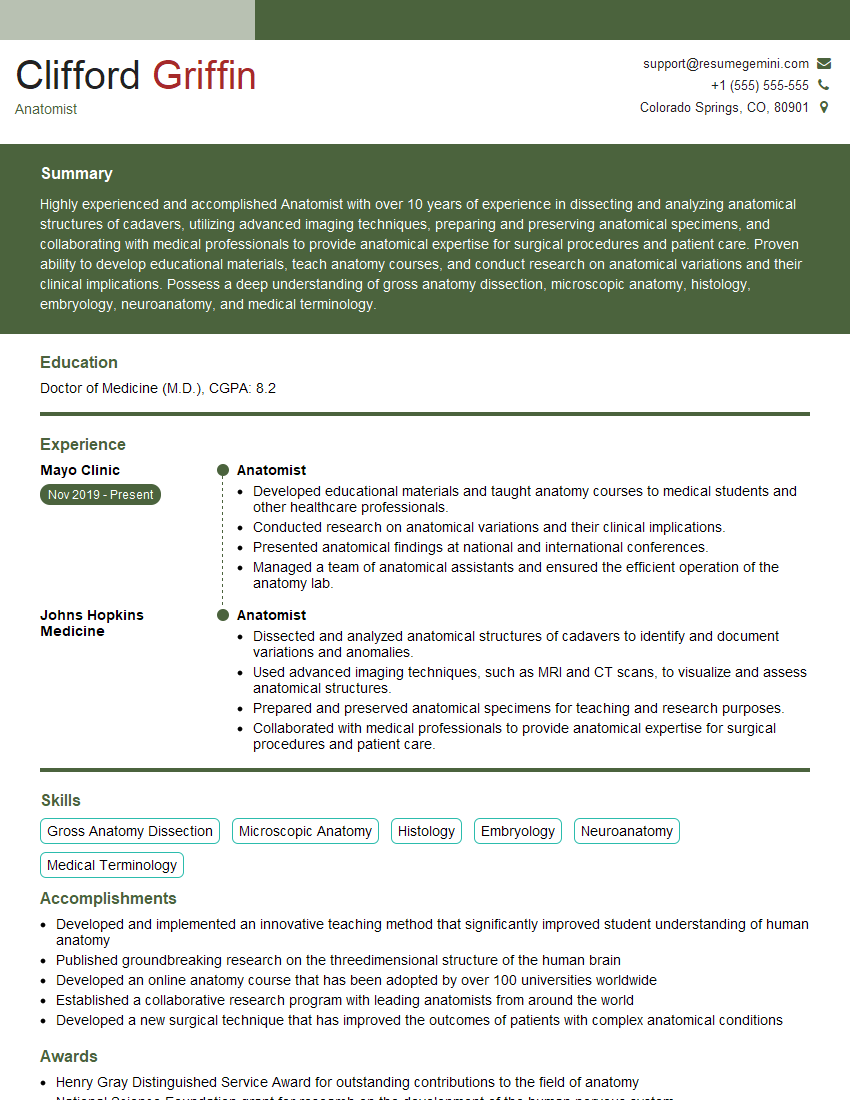
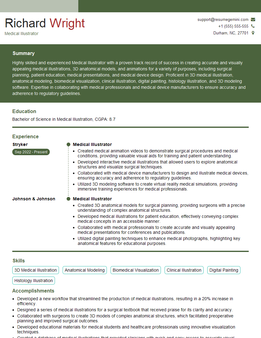
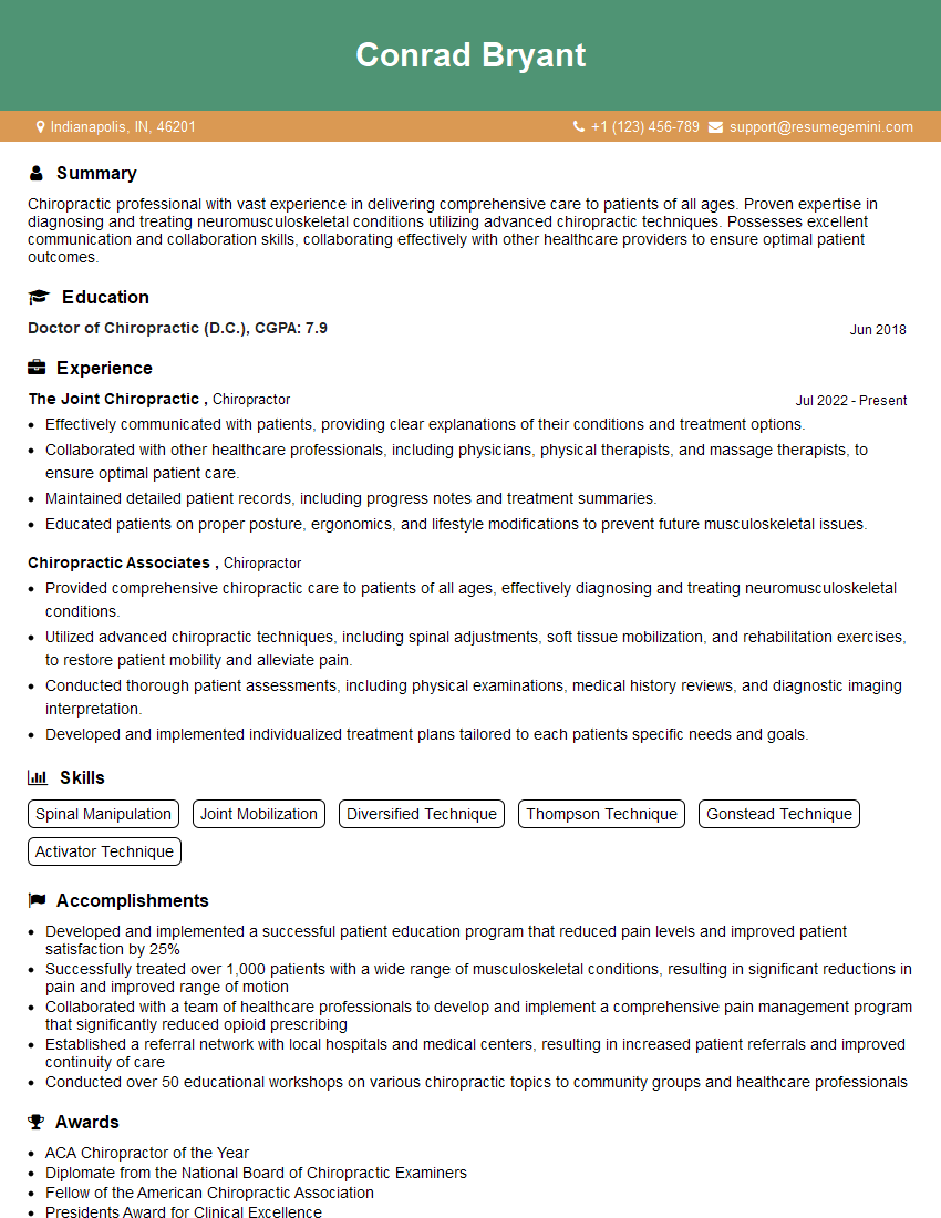
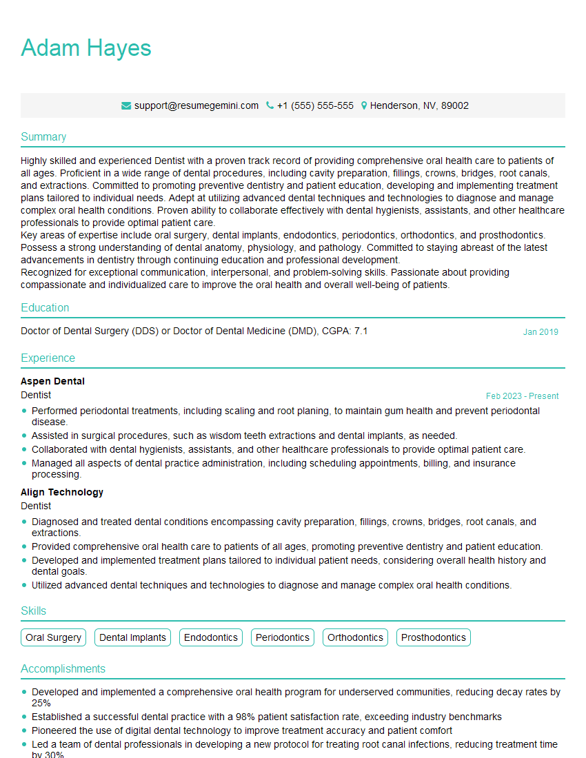
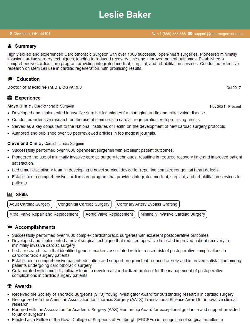
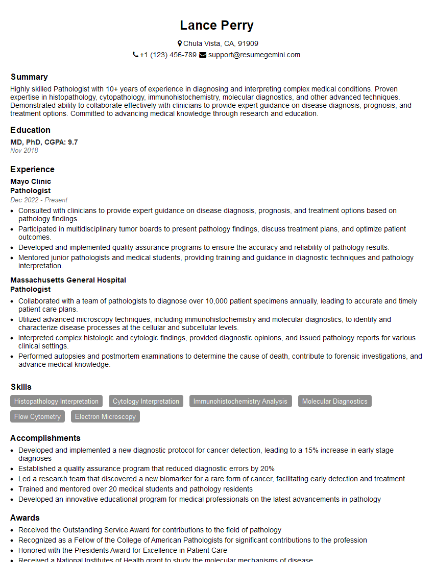
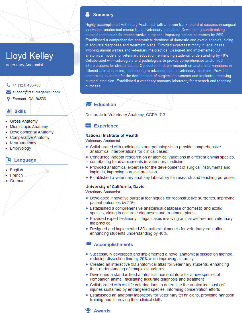
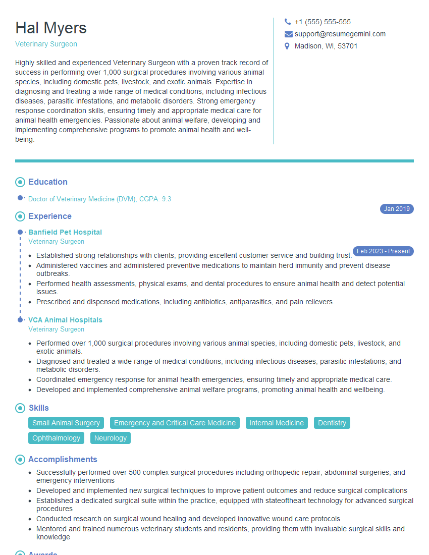
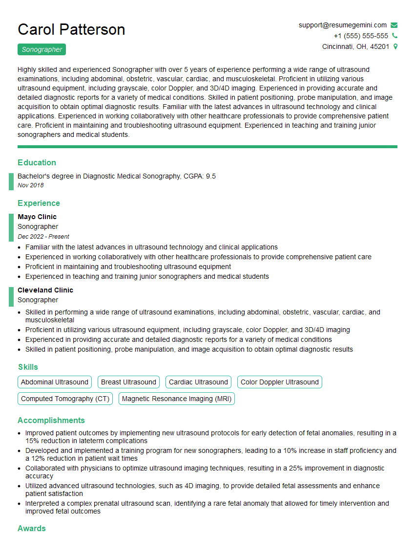
Mastering human and animal anatomy is crucial for career advancement in fields like medicine, veterinary science, research, and education. A strong understanding of this fundamental subject will significantly enhance your job prospects and allow you to contribute meaningfully to your chosen field. To maximize your chances of success, crafting a well-structured, ATS-friendly resume is paramount. ResumeGemini is a trusted resource to help you build a compelling and effective resume that highlights your skills and experience. ResumeGemini provides examples of resumes tailored to highlight a strong understanding of human and animal anatomy; review these examples to guide your own resume creation.
Explore more articles
Users Rating of Our Blogs
Share Your Experience
We value your feedback! Please rate our content and share your thoughts (optional).
What Readers Say About Our Blog
Hello,
We found issues with your domain’s email setup that may be sending your messages to spam or blocking them completely. InboxShield Mini shows you how to fix it in minutes — no tech skills required.
Scan your domain now for details: https://inboxshield-mini.com/
— Adam @ InboxShield Mini
Reply STOP to unsubscribe
Hi, are you owner of interviewgemini.com? What if I told you I could help you find extra time in your schedule, reconnect with leads you didn’t even realize you missed, and bring in more “I want to work with you” conversations, without increasing your ad spend or hiring a full-time employee?
All with a flexible, budget-friendly service that could easily pay for itself. Sounds good?
Would it be nice to jump on a quick 10-minute call so I can show you exactly how we make this work?
Best,
Hapei
Marketing Director
Hey, I know you’re the owner of interviewgemini.com. I’ll be quick.
Fundraising for your business is tough and time-consuming. We make it easier by guaranteeing two private investor meetings each month, for six months. No demos, no pitch events – just direct introductions to active investors matched to your startup.
If youR17;re raising, this could help you build real momentum. Want me to send more info?
Hi, I represent an SEO company that specialises in getting you AI citations and higher rankings on Google. I’d like to offer you a 100% free SEO audit for your website. Would you be interested?
Hi, I represent an SEO company that specialises in getting you AI citations and higher rankings on Google. I’d like to offer you a 100% free SEO audit for your website. Would you be interested?
good
