Preparation is the key to success in any interview. In this post, we’ll explore crucial Physical Assessment and Evaluation interview questions and equip you with strategies to craft impactful answers. Whether you’re a beginner or a pro, these tips will elevate your preparation.
Questions Asked in Physical Assessment and Evaluation Interview
Q 1. Describe the process of performing a neurological assessment.
A neurological assessment systematically evaluates the patient’s central and peripheral nervous systems. It’s crucial for detecting neurological deficits or disorders. The process typically involves several key steps:
- Mental Status Examination: Assessing level of consciousness (alert, drowsy, stuporous, comatose), orientation (person, place, time), memory, attention, and cognitive function. For example, I’d ask the patient their name, location, and the current date. Any confusion or disorientation would be noted.
- Cranial Nerves: Testing the 12 cranial nerves individually to assess their function. This involves testing vision (II), eye movements (III, IV, VI), facial sensation and movement (V, VII), hearing (VIII), swallowing (IX, X), shoulder elevation (XI), and tongue movement (XII). For instance, I’d test visual acuity using a Snellen chart and assess facial symmetry by asking the patient to smile and frown.
- Motor System: Evaluating muscle strength, tone, coordination, and involuntary movements. This includes assessing muscle bulk, range of motion, and presence of tremors or spasticity. I might ask the patient to perform rapid alternating movements or to walk heel-to-toe to assess coordination.
- Sensory System: Testing for sensation, including light touch, pain, temperature, and proprioception (sense of position). I would use a cotton swab to test light touch and a pinprick to assess pain sensation.
- Reflexes: Assessing deep tendon reflexes (e.g., biceps, triceps, patellar, Achilles) using a reflex hammer. The response is graded on a scale (e.g., 0-4+). An exaggerated reflex might suggest an upper motor neuron lesion.
- Cerebellar Function: Evaluating balance and coordination through tests like Romberg test (standing with eyes closed) and finger-to-nose test.
The findings are then synthesized to determine the overall neurological status of the patient. Any abnormalities are documented meticulously, highlighting their location, severity, and any potential causative factors.
Q 2. Explain the difference between objective and subjective data in physical assessment.
Objective and subjective data are two fundamental components of a thorough physical assessment. They differ significantly in their nature and how they’re obtained:
- Subjective Data: This is information provided by the patient, reflecting their personal experiences, feelings, and perceptions. It’s qualitative in nature and relies heavily on the patient’s reporting. Examples include pain levels (described as sharp, dull, aching), symptoms (e.g., nausea, dizziness), and medical history. A patient stating, “I have a sharp pain in my chest,” is subjective data.
- Objective Data: This is information gathered through direct observation, physical examination, and measurement. It’s quantitative and verifiable. Examples include vital signs (blood pressure, heart rate, temperature), physical findings (e.g., skin rash, abnormal lung sounds), and lab results. Observing a patient’s rapid respiratory rate of 30 breaths per minute is objective data.
Both types of data are crucial for a complete assessment. Subjective data provides context and the patient’s perspective, while objective data provides measurable evidence to support or refute the subjective information. Integrating both allows for a more accurate diagnosis and treatment plan.
Q 3. How do you assess a patient’s respiratory status?
Assessing respiratory status involves a multi-faceted approach, aiming to evaluate the effectiveness of gas exchange and the overall function of the respiratory system. I typically assess:
- Respiratory Rate and Rhythm: Observing the number of breaths per minute and their regularity. Tachypnea (rapid breathing), bradypnea (slow breathing), and apnea (absence of breathing) are important to note.
- Depth of Breathing: Assessing the volume of air exchanged with each breath. Shallow breathing may indicate respiratory distress or underlying pathology.
- Use of Accessory Muscles: Observing if the patient is using accessory muscles (e.g., neck muscles, intercostal muscles) to aid in breathing. This often suggests increased respiratory effort or distress.
- Breath Sounds: Auscultating the lungs using a stethoscope to detect normal or abnormal breath sounds. Crackles, wheezes, rhonchi, or diminished breath sounds can provide valuable clues about underlying lung conditions.
- Oxygen Saturation (SpO2): Measuring the percentage of oxygen in the blood using pulse oximetry. A low SpO2 indicates hypoxemia (low blood oxygen levels) which requires immediate attention.
- Work of Breathing: Observing the patient’s effort in breathing and noting any signs of distress like nasal flaring, retractions, or use of accessory muscles.
For example, a patient with pneumonia might exhibit increased respiratory rate, use of accessory muscles, crackles on auscultation, and decreased oxygen saturation.
Q 4. What are the key components of a cardiovascular assessment?
A comprehensive cardiovascular assessment aims to evaluate the heart’s structure and function. Key components include:
- Vital Signs: Measuring blood pressure (both systolic and diastolic), heart rate, and respiratory rate. Variations from the normal range can indicate underlying cardiovascular issues.
- Heart Rate and Rhythm: Assessing the rate and regularity of the heartbeat by palpating the radial pulse or auscultating the apical pulse. Arrhythmias (irregular heartbeats) can be detected.
- Blood Pressure: Measuring blood pressure using a sphygmomanometer to assess the pressure exerted by the blood against the arterial walls. Hypertension (high blood pressure) and hypotension (low blood pressure) are significant findings.
- Heart Sounds: Auscultating the heart using a stethoscope at various locations on the chest to identify normal heart sounds (S1 and S2) and any abnormal sounds such as murmurs, gallops, or rubs. Murmurs indicate turbulent blood flow, often indicative of valvular abnormalities.
- Peripheral Pulses: Assessing the strength and quality of peripheral pulses (e.g., radial, brachial, femoral, pedal) to evaluate blood flow to the extremities. Weak or absent pulses can signify peripheral artery disease.
- Inspection: Observing the patient for signs of edema (swelling), cyanosis (bluish discoloration), or jugular venous distention (JVD), which can suggest heart failure or other cardiovascular problems.
Careful interpretation of these findings helps in detecting conditions such as heart failure, valvular disease, arrhythmias, and hypertension.
Q 5. Describe your approach to assessing musculoskeletal injuries.
Assessing musculoskeletal injuries requires a systematic approach, integrating history, physical examination, and potentially imaging studies. My approach involves:
- History: Gathering detailed information about the mechanism of injury (e.g., fall, direct blow), onset of pain, location of pain, associated symptoms (e.g., swelling, deformity), and any previous injuries.
- Inspection: Observing the affected area for signs of deformity, swelling, bruising, or discoloration. Comparing the injured limb to the uninjured limb is essential.
- Palpation: Gently palpating the injured area to assess for tenderness, crepitus (a grating sound), muscle spasms, or bony deformities. This helps to localize the injury.
- Range of Motion (ROM): Assessing active and passive ROM of the affected joint. Limited ROM often indicates inflammation or injury to the joint or surrounding structures.
- Muscle Strength Testing: Evaluating muscle strength around the injured area to detect any weakness or paralysis.
- Neurovascular Assessment: Checking for distal pulses, skin color, temperature, sensation, and motor function to ensure adequate blood supply and nerve function. Changes in these indicate potential nerve or vascular compromise.
For example, if a patient presents with a suspected ankle sprain, I’d follow these steps to assess the extent of the injury, and may order x-rays to rule out fractures.
Q 6. How do you document your findings from a physical assessment?
Documentation of physical assessment findings is crucial for effective communication and continuity of care. I use a structured approach to ensure completeness and clarity:
- Patient Identification: Clearly identifying the patient with their name, date of birth, and medical record number.
- Date and Time: Recording the date and time of the assessment.
- Chief Complaint: Briefly stating the reason for the assessment.
- Subjective Data: Recording the patient’s statements, including symptoms, medical history, and current medications.
- Objective Data: Documenting all physical examination findings in a systematic and organized manner. Using specific terminology for describing findings is key (e.g., describing skin lesions as “erythematous macules” instead of “red spots”).
- Assessment: A concise summary of the clinician’s interpretation of the findings, indicating potential diagnoses or problems.
- Plan: Outlining the plan of care, including diagnostic tests, treatment plans, and follow-up appointments.
I use standardized formats and electronic health records (EHR) whenever possible to ensure consistency and accessibility. For example, I would document “Heart rate: 80 bpm, regular; Blood pressure: 120/80 mmHg; Lung sounds: clear to auscultation.” This level of detail is essential for proper documentation.
Q 7. Explain the importance of proper documentation in physical assessment.
Proper documentation in physical assessment is paramount for several reasons:
- Legal Protection: Detailed and accurate documentation serves as a legal record of the assessment, protecting both the patient and the healthcare provider. It can help resolve disputes or defend against malpractice claims.
- Continuity of Care: Comprehensive documentation allows healthcare providers to effectively communicate assessment findings and treatment plans, ensuring seamless transitions of care between different healthcare settings and providers.
- Improved Patient Care: Accurate documentation enhances the quality of patient care by providing a complete and reliable record of the patient’s condition, facilitating appropriate diagnosis and management.
- Research and Quality Improvement: Data from well-documented physical assessments contribute to research studies and quality improvement initiatives, leading to advancements in healthcare and improved patient outcomes. Aggregated data can track trends and aid in evidence-based decision-making.
- Reimbursement: Accurate documentation of services performed is essential for billing and reimbursement purposes.
In essence, thorough documentation not only meets legal and administrative requirements, but also directly impacts patient safety and the overall quality of healthcare delivery. Think of it as a crucial thread connecting various aspects of a patient’s journey, ensuring continuity and accuracy of their care.
Q 8. How do you adapt your assessment techniques for patients with different age groups?
Adapting assessment techniques to different age groups is crucial for accurate and effective evaluation. It requires understanding the developmental milestones and physiological changes associated with each stage of life.
- Infants (0-12 months): Assessments focus heavily on observation. We look at reflexes (Moro, rooting, sucking), response to stimuli, and vital signs. Communication is primarily nonverbal, relying on soothing techniques and careful handling. For example, assessing heart rate in an infant might involve auscultation while the baby is calmly held by a parent.
- Children (1-12 years): Communication becomes more interactive, incorporating play and simple language. Assessments involve age-appropriate games to evaluate cognitive function and motor skills. Explaining procedures in simple terms is key to building trust. For example, demonstrating a stethoscope on a toy before using it on the child builds rapport.
- Adolescents (13-18 years): Privacy and respect for autonomy are paramount. Open communication is encouraged, addressing concerns and respecting their perspectives. It’s important to involve them in the decision-making process about their care. For example, involving them in deciding which parts of the exam they want to do first builds their confidence in you and makes them feel respected.
- Adults (19-64 years): Standard physical assessment techniques are typically applicable. Clear communication, informed consent, and respect for their privacy are crucial. Adapting to physical limitations (e.g., using alternative positions for examination) is important.
- Older Adults (65+ years): Consideration of age-related changes (decreased vision, hearing loss, decreased mobility) is essential. Adjust the pace of the examination, provide ample time for questions, and use assistive devices as needed. For example, using a larger print font or speaking slowly helps ensure clarity during communication.
In summary, flexibility and adaptability are keys to performing accurate assessments across the lifespan. Knowing the specific needs of each age group and adjusting our approach accordingly ensures we gather the most comprehensive and reliable data possible.
Q 9. What are the common signs and symptoms of a stroke and how would you assess for them?
Stroke, or cerebrovascular accident (CVA), is a medical emergency caused by interruption of blood flow to the brain. Recognizing the signs and symptoms quickly is critical for timely intervention and improved outcomes. The most common signs and symptoms are often remembered by the acronym FAST:
- Facial drooping: Ask the person to smile. Does one side of the face droop?
- Arm weakness: Ask the person to raise both arms. Does one arm drift downward?
- Speech difficulty: Ask the person to repeat a simple phrase. Is their speech slurred or strange?
- Time: If you observe any of these signs, call emergency services immediately.
Beyond FAST, other potential assessment findings include:
- Sudden severe headache (often described as the “worst headache of their life”)
- Confusion, disorientation, or difficulty understanding
- Loss of balance or coordination
- Vision changes (blurred vision, double vision)
- Numbness or weakness on one side of the body
- Difficulty swallowing
Assessment involves a thorough neurological exam, including checking for:
- Level of consciousness
- Pupillary response
- Cranial nerve function
- Motor strength and coordination
- Sensory function
- Reflexes
It’s vital to act quickly. The time sensitivity is due to the potential for irreversible brain damage; early treatment is key to improving outcomes.
Q 10. How do you differentiate between different types of pain?
Differentiating between types of pain requires a comprehensive approach, going beyond simply asking the patient where it hurts. We need to understand the characteristics of the pain to determine its potential cause. Pain is subjective, so the patient’s description is crucial.
Key characteristics to assess include:
- Location: Where exactly is the pain located? Is it localized or radiating?
- Quality: How would the patient describe the pain? (e.g., sharp, dull, aching, burning, throbbing, stabbing)
- Intensity: How severe is the pain on a scale of 0-10 (or using a visual analog scale)?
- Timing: When did the pain start? Is it constant, intermittent, or does it come and go? How long does it last?
- Aggravating and relieving factors: What makes the pain worse or better? This can offer valuable clues about its potential source.
- Associated symptoms: Are there any other symptoms present, such as nausea, vomiting, fever, or neurological changes?
Examples of different pain types and their characteristics:
- Nociceptive pain: This arises from damage to tissues and is usually well-localized. Examples include pain from a cut or a sprain. It often responds well to standard pain relievers.
- Neuropathic pain: This results from damage to the nerves themselves. It is often described as burning, tingling, or shooting pain, and is often difficult to treat. Examples include diabetic neuropathy or post-herpetic neuralgia.
- Visceral pain: This originates from internal organs and is often poorly localized, diffuse, and accompanied by other symptoms like nausea or vomiting. Examples include appendicitis or pancreatitis.
- Referred pain: This pain is felt in a location different from its actual source. For example, heart attack pain is often felt in the left arm or jaw.
Using a structured approach and carefully considering these factors helps in differentiating types of pain and guiding appropriate treatment strategies.
Q 11. Describe the techniques used to assess skin integrity.
Assessing skin integrity involves a thorough examination to identify any abnormalities or impairments. This evaluation is crucial for preventing and managing pressure injuries, infections, and other skin-related complications. The assessment should be systematic and include:
- Visual Inspection: Observe the skin’s color, texture, moisture, temperature, and presence of any lesions, wounds, or discoloration. Pay close attention to areas prone to pressure injuries, like bony prominences. Look for signs of inflammation like redness, swelling, or warmth.
- Palpation: Gently palpate the skin to assess its turgor (elasticity), temperature, and the presence of any edema (swelling), masses, or tenderness. Assess for any skin breakdown and its depth using a Braden scale to classify the risk.
- Wound Assessment (if present): If a wound or lesion is present, document its location, size (length, width, depth), appearance (color, presence of exudate, necrotic tissue), and surrounding skin condition. This helps monitor progress and treatment effectiveness.
- Documentation: Thorough and accurate documentation of the assessment findings, including photographs if appropriate, is vital for tracking changes over time and ensuring continuity of care. The documentation should be specific and detailed. For example, instead of writing “skin tear,” it would be better to describe: “2cm x 1cm skin tear, located on the left elbow, with serosanguineous drainage.”
A systematic approach to skin assessment is essential for early detection of potential problems and the implementation of preventive measures. Regular assessment, especially for patients at high risk (e.g., bedridden patients, those with impaired mobility, or those with diabetes), is critical to preserving skin integrity.
Q 12. How do you assess a patient’s level of consciousness?
Assessing a patient’s level of consciousness (LOC) is a fundamental aspect of neurological examination. It helps determine the patient’s alertness and responsiveness to stimuli. Various scales are used for this purpose, the most common being the Glasgow Coma Scale (GCS).
The GCS assesses three aspects:
- Eye opening: Spontaneous (4 points), to speech (3 points), to pain (2 points), none (1 point)
- Verbal response: Oriented (5 points), confused conversation (4 points), inappropriate words (3 points), incomprehensible sounds (2 points), none (1 point)
- Motor response: Obeys commands (6 points), localizes pain (5 points), withdraws from pain (4 points), flexor posturing (decorticate, 3 points), extensor posturing (decerebrate, 2 points), none (1 point)
The scores are added together; a total score of 15 indicates full alertness, while a score of 3 represents deep coma. Other methods for assessing LOC include:
- AVPU scale: Alert, Verbal stimuli response, Painful stimuli response, Unresponsive
- Observation: Note the patient’s spontaneous movements, responsiveness to questions, and overall alertness.
It is important to note that the GCS is not solely used to determine the level of consciousness, but rather the severity of neurological damage as determined by the level of consciousness in the assessment.
Q 13. What are the different scales used to assess pain?
Several scales are used to assess pain, each with its strengths and limitations. The choice of scale depends on the patient’s cognitive abilities and communication skills.
- Numeric Rating Scale (NRS): A 0-10 scale where 0 represents no pain and 10 represents the worst imaginable pain. Simple and widely used, but it relies on the patient’s ability to understand numbers.
- Visual Analog Scale (VAS): A 10cm line where the patient marks their pain level. Useful for patients with language barriers or cognitive impairment, but interpretation can be subjective.
- Wong-Baker FACES Pain Rating Scale: Uses cartoon faces with different expressions representing various pain levels. Ideal for children or patients with cognitive deficits who cannot understand numbers.
- Verbal Descriptor Scale (VDS): Provides a list of words describing pain intensity (e.g., mild, moderate, severe, unbearable). Useful for patients who can verbally communicate but might struggle with numerical scales.
- FLACC Scale: Face, Legs, Activity, Cry, Consolability. Uses observations of nonverbal cues to assess pain in infants and young children who cannot self-report. It’s based on observation of the patient.
The most appropriate scale should be selected based on the individual patient’s needs and abilities. A combination of scales can be used for a more comprehensive assessment.
Q 14. How do you assess a patient’s cognitive function?
Assessing cognitive function involves evaluating various aspects of mental processing, including attention, memory, language, and executive function. The approach depends on the patient’s age, medical history, and presenting concerns. Several standardized tests are available, including the Mini-Mental State Examination (MMSE) and the Montreal Cognitive Assessment (MoCA).
Assessment may include:
- Orientation: Assess the patient’s awareness of time, place, and person (who they are, where they are, what the date is).
- Attention and Concentration: Test attention span using tasks such as serial 7s subtraction or spelling ‘WORLD’ backwards.
- Memory: Evaluate both short-term and long-term memory by asking the patient to recall information immediately after it is presented and later in the assessment.
- Language: Assess fluency, comprehension, repetition, and naming abilities.
- Executive Function: Assess abilities such as planning, problem-solving, and abstract thinking. This may involve tasks like drawing a clock or interpreting proverbs.
- Insight and Judgment: Assess the patient’s awareness of their cognitive deficits and their ability to make sound decisions.
Observation of the patient’s behavior during the assessment is also crucial. Changes in personality, mood, and behavior can be significant indicators of cognitive impairment. A combination of standardized tests and observational assessment is often necessary for a thorough evaluation.
Remember to consider the cultural background and educational level of the patient when interpreting results. For instance, a patient with limited education might score lower on certain tests even if their cognitive function is intact.
Q 15. What are the early signs of infection and how would you assess for them?
Early signs of infection, often called prodromal symptoms, can be subtle and vary depending on the location and type of infection. They often manifest as a systemic inflammatory response. Assessing for them requires a systematic approach focusing on vital signs and specific symptoms.
- Fever: Elevated body temperature (oral temperature > 100.4°F or 38°C) is a common indicator. I would assess this using a thermometer, ensuring proper technique and considering factors like time of day and recent activity.
- Tachycardia: An increased heart rate (above the normal range for the patient’s age and condition) suggests the body is working harder to fight the infection. I would assess this using a stethoscope and counting heart beats for a full minute.
- Tachypnea: Increased respiratory rate (faster than the normal range) indicates the body’s effort to increase oxygen intake. I would assess this by observing the patient’s breathing rate for one minute.
- Changes in White Blood Cell Count (WBC): Although not directly assessed during a physical examination, a complete blood count (CBC) revealing elevated WBCs would strongly support the presence of an infection. This is a laboratory finding, requiring a blood sample.
- Malaise/Fatigue: Generalized feeling of discomfort, weakness, and tiredness are often early indicators. This is assessed through direct questioning of the patient, paying attention to their description of their energy levels.
- Localized Symptoms: Depending on the site of infection, there might be localized redness, warmth, swelling, pain, or purulent drainage (pus). This would involve a careful visual inspection and palpation of the affected area.
For example, a patient presenting with a low-grade fever, fatigue, and a slightly reddened, tender throat would raise suspicion for a developing pharyngitis. Combining these clinical findings with a thorough history would help establish a diagnosis.
Career Expert Tips:
- Ace those interviews! Prepare effectively by reviewing the Top 50 Most Common Interview Questions on ResumeGemini.
- Navigate your job search with confidence! Explore a wide range of Career Tips on ResumeGemini. Learn about common challenges and recommendations to overcome them.
- Craft the perfect resume! Master the Art of Resume Writing with ResumeGemini’s guide. Showcase your unique qualifications and achievements effectively.
- Don’t miss out on holiday savings! Build your dream resume with ResumeGemini’s ATS optimized templates.
Q 16. Describe your approach to assessing a patient’s nutritional status.
Assessing a patient’s nutritional status is crucial for overall health evaluation. It’s a multi-faceted assessment encompassing several key areas.
- Anthropometric Measurements: This involves measuring height, weight, BMI (Body Mass Index), waist circumference, and skinfold thickness. These measures help determine overall body composition and identify potential malnutrition or obesity.
- Dietary History: A detailed dietary recall (24-hour or multiple-day) or food frequency questionnaire is essential. I would inquire about the types of food consumed, portion sizes, and frequency. This allows me to assess the balance of macronutrients (carbohydrates, proteins, fats) and micronutrients (vitamins and minerals).
- Laboratory Tests: Serum albumin, prealbumin, transferrin, and total lymphocyte count are blood tests that reflect protein status, reflecting nutritional adequacy. These tests provide objective data to supplement clinical assessment.
- Physical Examination: I would look for signs of vitamin deficiencies (e.g., pallor indicating anemia, changes in skin integrity, or muscle wasting). I would also assess the patient’s energy levels and activity tolerance, which can be markedly affected by nutritional deficiencies.
- Patient History: Inquiring about factors such as appetite changes, difficulty chewing or swallowing, recent weight changes, use of medications, and socioeconomic factors affecting food access is important.
For instance, a patient with a low BMI, pale skin, and reporting a poor appetite alongside a history of decreased food intake would prompt further investigation of potential malnutrition.
Q 17. How do you assess a patient’s fluid balance?
Assessing fluid balance requires a comprehensive approach, combining the patient’s history with a physical examination and, if necessary, laboratory tests.
- Intake and Output (I&O): This is a fundamental aspect. I would meticulously record the patient’s fluid intake (oral fluids, intravenous fluids) and output (urine, stool, vomitus, drainage). Discrepancies can point towards fluid imbalance.
- Daily Weights: Weighing the patient daily at the same time of day provides an objective measure of fluid changes. A significant weight gain (usually > 2 pounds in a day) often points towards fluid retention, whereas significant weight loss suggests dehydration.
- Vital Signs: Changes in blood pressure (hypotension in dehydration, hypertension in fluid overload), heart rate (tachycardia in dehydration), and respiratory rate (tachypnea in pulmonary edema) can be indicative of fluid imbalances.
- Physical Examination: I would assess skin turgor (checking skin elasticity by gently pinching the skin—tenting signifies dehydration), mucous membrane moisture (dry mucous membranes indicate dehydration), and edema (swelling, often in dependent areas, indicates fluid overload). Jugular venous distention (JVD) can suggest fluid overload.
- Laboratory Tests: Electrolytes (sodium, potassium), blood urea nitrogen (BUN), and creatinine provide additional objective data on fluid balance and kidney function.
A patient with decreased urine output, dry mucous membranes, and orthostatic hypotension (drop in blood pressure upon standing) might be severely dehydrated and require immediate intervention.
Q 18. What are the common signs and symptoms of dehydration and how would you assess for them?
Dehydration is a serious condition resulting from a fluid deficiency in the body. Signs and symptoms can range from mild to severe depending on the severity of fluid loss.
- Mild Dehydration: Symptoms often include thirst, dry mouth, slightly decreased urine output, and mild fatigue. I would assess these through direct patient questioning and observing the color and concentration of urine.
- Moderate Dehydration: In addition to the mild symptoms, moderate dehydration may present with dry skin and mucous membranes, sunken eyes, low blood pressure, increased heart rate, and dizziness. Assessment involves observing these physical findings, palpating skin turgor, and measuring blood pressure and heart rate.
- Severe Dehydration: This is a life-threatening condition characterized by extreme thirst, very little to no urine output, very low blood pressure, rapid and weak pulse, rapid breathing, confusion, and even loss of consciousness. This requires immediate medical intervention, including intravenous fluids. Assessment requires comprehensive evaluation of vital signs, mental status, and careful observation of the patient’s overall condition.
For example, a child presenting with lethargy, sunken eyes, and no tears upon crying would strongly suggest severe dehydration requiring immediate medical attention.
Q 19. How would you assess for signs of potential abuse or neglect?
Assessing for potential abuse or neglect requires a sensitive and systematic approach, combining observation with direct questioning and utilizing appropriate documentation.
- Physical Examination: Look for unexplained bruises, burns (especially in patterns), fractures, or other injuries, paying close attention to inconsistencies between the explanation and the injury’s appearance. Note the location, size, and shape of the injuries.
- History Taking: Inquire about the circumstances surrounding the injuries or concerning situations. Pay attention to any inconsistencies, hesitancy in answering, or discrepancies in the explanation provided by the patient or caregiver. Maintain a non-judgmental and empathetic approach.
- Behavioral Observations: Observe the patient’s behavior and interactions with caregivers. Look for fear, anxiety, withdrawal, or unusual compliance or passivity. Note any signs of neglect, such as poor hygiene, malnutrition, or inappropriate clothing for the environment.
- Documentation: Meticulously document all observations, including detailed descriptions of injuries, inconsistencies in the reported history, and the patient’s behavior. Use objective and factual language, avoiding subjective interpretations.
- Mandatory Reporting: In situations where abuse or neglect is suspected, it’s crucial to follow mandatory reporting laws and regulations in the jurisdiction. This usually involves contacting the appropriate authorities, such as child protective services or adult protective services.
If a patient presents with unexplained bruises in various stages of healing and is reluctant to discuss their home environment, it would be necessary to undertake further investigations, ensuring patient safety and compliance with reporting regulations.
Q 20. Describe your experience with using different diagnostic equipment in physical assessment (e.g., stethoscope, spirometer).
I have extensive experience using various diagnostic equipment in physical assessment. My proficiency includes:
- Stethoscope: I utilize a stethoscope daily to auscultate heart sounds (identifying murmurs, gallops, arrhythmias), lung sounds (detecting wheezes, crackles, rhonchi), and bowel sounds (assessing gut motility). Proper technique, including correct placement and listening for distinct sounds, is crucial for accurate interpretation.
- Sphygmomanometer: Precise blood pressure measurement is essential. I am proficient in using a sphygmomanometer to accurately assess systolic and diastolic pressures, and I understand the importance of proper cuff size selection and technique to avoid errors.
- Spiometer: I am comfortable using a spirometer to assess lung function, including forced expiratory volume (FEV1) and forced vital capacity (FVC). I am aware of the importance of patient instruction and proper technique for obtaining reliable measurements.
- Other Equipment: My experience also extends to the use of other equipment like ophthalmoscopes (for eye examination), otoscopes (for ear examination), reflex hammers (for neurological assessment), and scales (for weighing patients).
For example, while assessing a patient with shortness of breath, I would use a stethoscope to listen for adventitious lung sounds, and a spirometer to objectively assess lung function. The combination of these methods yields a more complete picture of the patient’s respiratory status.
Q 21. Explain the importance of obtaining a complete medical history before conducting a physical assessment.
Obtaining a complete medical history before conducting a physical assessment is paramount because it provides crucial context for interpreting physical findings. The physical exam is only one piece of the puzzle. The history guides the assessment and helps to focus on areas needing specific attention.
- Directing the Physical Exam: The history helps to prioritize areas for examination. For example, a patient complaining of chest pain will necessitate a thorough cardiovascular assessment, whereas a patient with abdominal pain will require a focused abdominal exam.
- Identifying Risk Factors: The history helps identify potential risk factors for disease. Knowledge of smoking history, family history of heart disease, or exposure to infectious agents all significantly impact interpretation of physical findings.
- Understanding Symptoms: The history provides details about the onset, duration, character, location, and associated symptoms, allowing for a comprehensive understanding of the patient’s complaint and better discrimination between possible diagnoses.
- Developing a Differential Diagnosis: A complete history helps narrow down possible diagnoses, leading to a more efficient and targeted physical exam. This avoids unnecessary examinations and ensures relevant aspects are emphasized.
- Establishing Baseline: A thorough history establishes a baseline for future comparisons. It allows for tracking of changes in the patient’s condition over time, essential for managing chronic conditions and assessing the effectiveness of treatment.
For instance, a patient reporting a cough for three weeks, with associated night sweats and weight loss, would immediately suggest the need for a more detailed respiratory examination than a patient reporting a cough of only a few days’ duration.
Q 22. How do you prioritize your assessment based on patient acuity?
Prioritizing a physical assessment based on patient acuity involves a systematic approach focusing on the most immediate and life-threatening issues first. Think of it like a triage system in an emergency room – you address the most critical needs before moving to less urgent concerns.
- ABCDE approach: This is a fundamental principle. We prioritize Airway, Breathing, Circulation, Disability (neurological status), and Exposure (environmental factors). If a patient is having difficulty breathing (B), that takes precedence over assessing their skin integrity (E).
- Urgent vs. Non-Urgent: Chest pain, severe bleeding, altered mental status, and respiratory distress are examples of urgent findings demanding immediate attention. Less urgent issues include rashes, mild pain, or a chronic health problem needing routine monitoring.
- Patient Presentation: The patient’s overall appearance can provide important clues. A patient in obvious distress or with signs of shock will require immediate assessment of vital signs and interventions.
For instance, a patient presenting with shortness of breath and cyanosis (bluish skin discoloration) will have their airway and breathing assessed immediately, even before detailed questioning about their medical history. This ensures prompt identification and management of life-threatening conditions.
Q 23. How do you handle unexpected findings during a physical assessment?
Unexpected findings during a physical assessment require a calm, systematic approach. The key is to remain objective, document thoroughly, and escalate concerns appropriately.
- Re-assess: First, I would re-assess the finding to confirm its accuracy. Is this a true finding, or a misinterpretation?
- Explore Related Symptoms: Are there other symptoms that could explain this finding? For instance, if I find an unexpected heart murmur, I would look for signs of heart failure, such as edema or shortness of breath.
- Differential Diagnosis: I’d mentally generate a differential diagnosis—a list of possible causes—for the unexpected finding. This will guide further investigation and appropriate testing.
- Consult and Document: I would promptly consult with a supervising physician or other member of the healthcare team, sharing my observations and proposed differential diagnosis. All findings, including unexpected ones, should be meticulously documented in the patient’s medical record.
- Patient Education: If appropriate, I would communicate the findings and their implications to the patient in a clear and empathetic manner.
For example, if I discovered an unexpected lump during a breast examination, I wouldn’t panic. Instead, I’d carefully palpate the lump, noting its size, consistency, and mobility, and then immediately discuss it with the physician to determine appropriate next steps, such as ordering a mammogram or ultrasound.
Q 24. How do you communicate your assessment findings to other members of the healthcare team?
Clear and concise communication is vital in healthcare. I use a standardized approach, ensuring accuracy and completeness in conveying assessment findings.
- SBAR framework: This is a structured method that involves Situation (brief description), Background (relevant history), Assessment (my findings), and Recommendation (suggested actions). This allows for efficient and accurate communication of critical information.
- Documentation: I maintain comprehensive, accurate, and timely documentation in the electronic health record (EHR) to ensure all relevant findings are recorded and readily available to the healthcare team.
- Verbal Communication: During handoffs or team meetings, I present findings clearly and professionally, using precise medical terminology while ensuring it’s understandable to the audience.
- Collaboration: I actively participate in team discussions, offering my insights and collaborating with other healthcare professionals to ensure a holistic approach to patient care.
For instance, using SBAR, I might report: ‘Situation: Patient presents with decreased breath sounds in the right lower lobe. Background: 80-year-old female with history of COPD. Assessment: Suspected pneumonia based on auscultation and vital signs. Recommendation: Chest X-ray and antibiotics.’
Q 25. Describe a situation where you had to modify your assessment approach due to a patient’s limitations.
I have had to modify my assessment approach many times due to patient limitations. Adaptability and creativity are crucial skills in physical assessment.
For instance, I assessed a patient with severe rheumatoid arthritis, who had limited mobility and significant pain. I could not perform a full range-of-motion assessment in the traditional manner. Instead, I focused on passively assessing range of motion, noting any resistance or pain, and observing any joint deformities. I also adjusted my approach to palpation, performing it gently and with sensitivity to minimize discomfort. I adapted my communication to be mindful of the patient’s pain and frustration, ensuring a compassionate environment throughout the exam. I emphasized active listening to understand their experience and perspectives.
Q 26. How do you ensure patient comfort and safety during a physical assessment?
Patient comfort and safety are paramount during a physical assessment. I employ several strategies to ensure both.
- Privacy and Dignity: I always ensure privacy by performing the assessment in a private room and explaining each step before proceeding. I ask permission to touch, providing a rationale. I use drapes appropriately to preserve modesty.
- Pain Management: I assess for pain at the beginning and throughout the exam. If a patient expresses discomfort, I adjust the exam accordingly, using alternative techniques or offering pain relief as appropriate.
- Infection Control: I maintain strict adherence to infection control practices, including proper hand hygiene, use of gloves, and appropriate disposal of waste.
- Positioning: I assist patients with comfortable positioning during the assessment, helping them move slowly to avoid falls or injury.
- Safety Precautions: If a patient is at risk of falling, I take precautions such as having them sit or lie down, offering assistance, and ensuring the environment is safe.
For example, when assessing an elderly patient with balance problems, I would ensure they are seated safely and make sure there’s a chair close by in case they feel faint or dizzy. I would also explain each step clearly and slowly, offering rest periods as needed.
Q 27. Explain your understanding of legal and ethical considerations related to physical assessment.
Legal and ethical considerations are fundamental to physical assessment. My practice adheres to strict professional guidelines.
- Consent: I obtain informed consent from the patient before commencing the assessment. This includes explaining the purpose, procedure, and potential risks and benefits.
- Confidentiality: Patient information gathered during the assessment is strictly confidential and protected according to HIPAA regulations and other relevant laws.
- Professional Boundaries: I maintain professional boundaries, avoiding any behavior that could be interpreted as inappropriate or unprofessional.
- Scope of Practice: I only perform assessments within my scope of practice, referring complex cases or findings outside my expertise to qualified healthcare professionals.
- Documentation: Accurate and thorough documentation protects both the patient and the healthcare provider from legal liability.
For instance, I would never perform a pelvic exam on a patient without their explicit consent, and I would ensure that all documentation is objective and factually accurate to avoid misinterpretations or legal challenges.
Q 28. How do you stay updated with current best practices in physical assessment and evaluation?
Staying updated with current best practices in physical assessment and evaluation is a continuous process. I utilize several strategies to ensure my knowledge remains current.
- Professional Organizations: I am a member of relevant professional organizations (e.g., American Academy of Nursing, etc.) that provide access to continuing education resources, journals, and conferences.
- Continuing Education: I actively participate in continuing education courses, workshops, and conferences related to physical assessment and evaluation.
- Peer Learning: I engage in peer learning opportunities with colleagues and attend interprofessional educational sessions.
- Professional Journals: I regularly read relevant professional journals and research articles to stay abreast of the latest research and advancements.
- Online Resources: I utilize reliable online resources and databases to access the latest guidelines, protocols, and best practices.
For example, I regularly review updated guidelines for assessing cardiovascular risk factors or changes to techniques for performing neurological exams, ensuring I’m consistently using the most accurate and effective methods.
Key Topics to Learn for Physical Assessment and Evaluation Interview
- Musculoskeletal System Assessment: Understanding range of motion, muscle strength testing, and palpation techniques. Practical application includes accurately documenting findings and differentiating between normal and abnormal presentations.
- Neurological Assessment: Mastering techniques for evaluating reflexes, coordination, balance, and sensory function. Practical application involves interpreting neurological findings to identify potential pathologies and inform treatment plans.
- Cardiopulmonary Assessment: Learning how to assess vital signs, auscultate heart and lung sounds, and interpret electrocardiograms (ECGs). Practical application includes recognizing signs of cardiac and respiratory distress and initiating appropriate interventions.
- Integumentary System Assessment: Developing proficiency in inspecting and palpating the skin for lesions, wounds, and other abnormalities. Practical application includes accurately documenting skin findings and understanding their significance.
- Documentation and Reporting: Understanding the importance of clear, concise, and accurate documentation of assessment findings. Practical application involves using standardized terminology and formatting to ensure effective communication with healthcare professionals.
- Ethical Considerations and Legal Implications: Understanding professional responsibilities, patient confidentiality, and legal aspects related to physical assessment and evaluation. Practical application includes adhering to ethical guidelines and maintaining patient privacy.
- Problem-Solving and Critical Thinking: Developing the ability to analyze assessment data, identify potential problems, and develop appropriate interventions. Practical application includes formulating differential diagnoses and collaborating with other healthcare professionals.
Next Steps
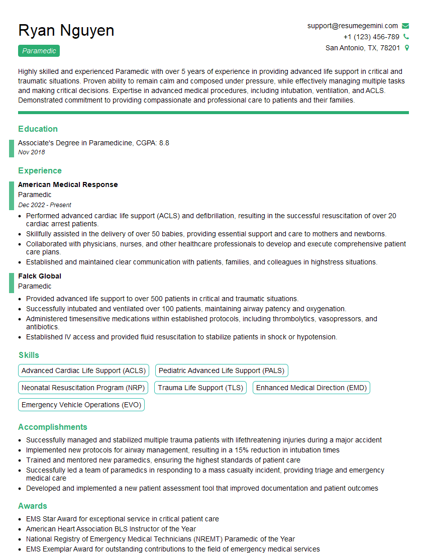
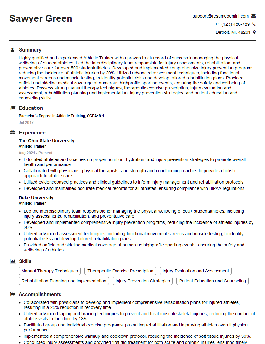
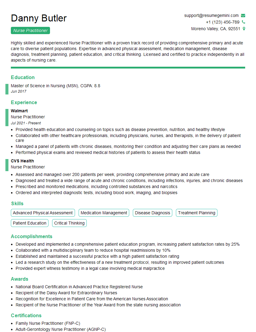
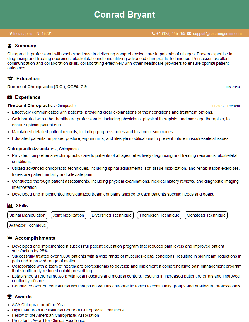
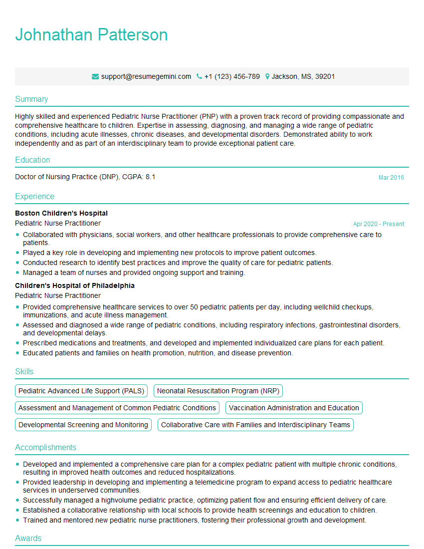
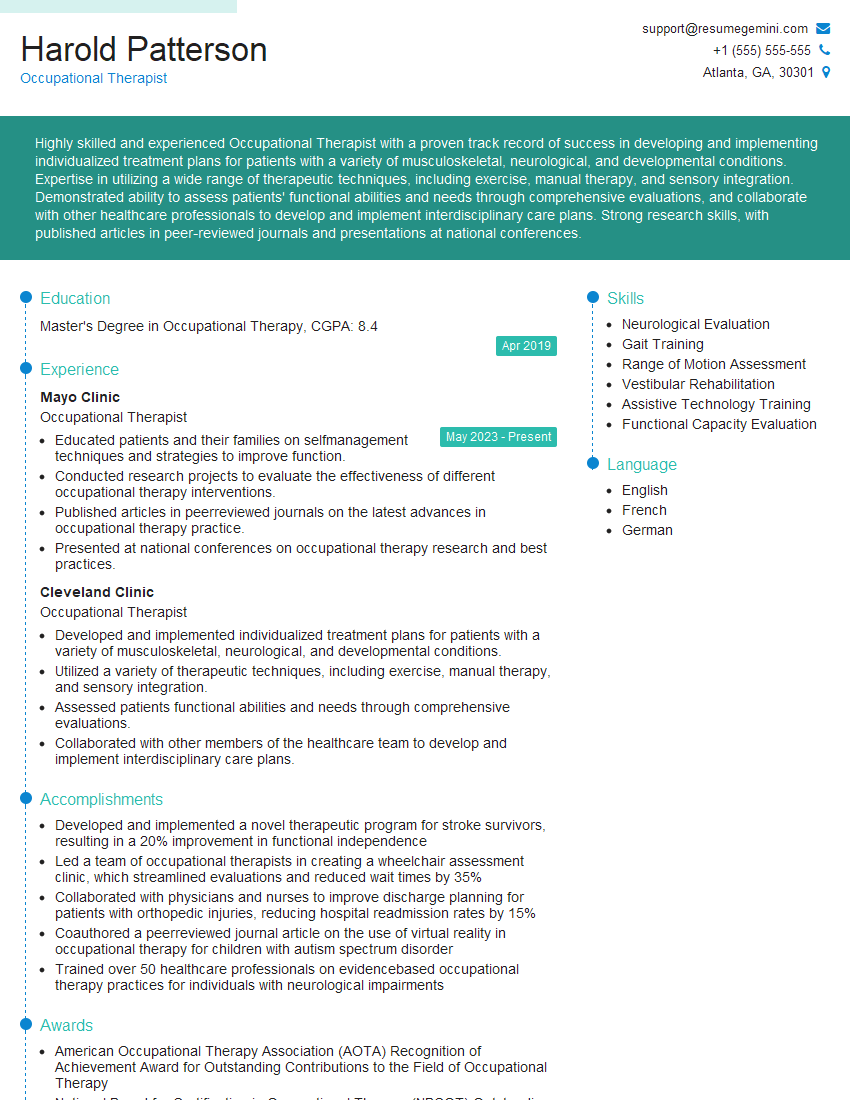
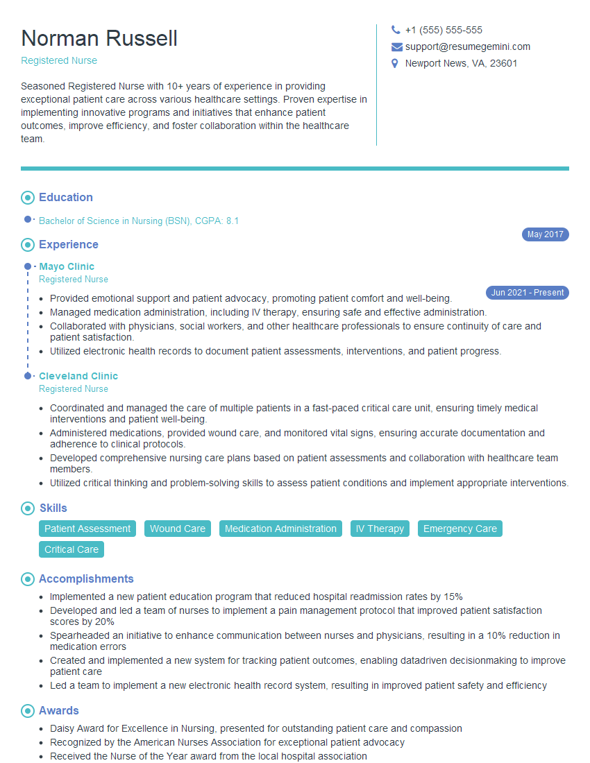
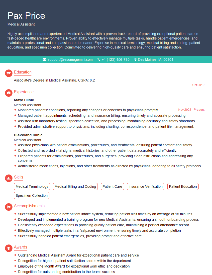
Mastering Physical Assessment and Evaluation is crucial for career advancement in healthcare, opening doors to specialized roles and increased earning potential. A strong resume is your key to unlocking these opportunities. Creating an ATS-friendly resume significantly improves your chances of getting noticed by recruiters. We strongly recommend using ResumeGemini to build a professional and impactful resume that highlights your skills and experience. ResumeGemini provides examples of resumes tailored to Physical Assessment and Evaluation roles to guide you through the process. Invest time in crafting a compelling resume – it’s your first impression and a crucial step in your career journey.
Explore more articles
Users Rating of Our Blogs
Share Your Experience
We value your feedback! Please rate our content and share your thoughts (optional).
What Readers Say About Our Blog
Hello,
We found issues with your domain’s email setup that may be sending your messages to spam or blocking them completely. InboxShield Mini shows you how to fix it in minutes — no tech skills required.
Scan your domain now for details: https://inboxshield-mini.com/
— Adam @ InboxShield Mini
Reply STOP to unsubscribe
Hi, are you owner of interviewgemini.com? What if I told you I could help you find extra time in your schedule, reconnect with leads you didn’t even realize you missed, and bring in more “I want to work with you” conversations, without increasing your ad spend or hiring a full-time employee?
All with a flexible, budget-friendly service that could easily pay for itself. Sounds good?
Would it be nice to jump on a quick 10-minute call so I can show you exactly how we make this work?
Best,
Hapei
Marketing Director
Hey, I know you’re the owner of interviewgemini.com. I’ll be quick.
Fundraising for your business is tough and time-consuming. We make it easier by guaranteeing two private investor meetings each month, for six months. No demos, no pitch events – just direct introductions to active investors matched to your startup.
If youR17;re raising, this could help you build real momentum. Want me to send more info?
Hi, I represent an SEO company that specialises in getting you AI citations and higher rankings on Google. I’d like to offer you a 100% free SEO audit for your website. Would you be interested?
Hi, I represent an SEO company that specialises in getting you AI citations and higher rankings on Google. I’d like to offer you a 100% free SEO audit for your website. Would you be interested?
good
