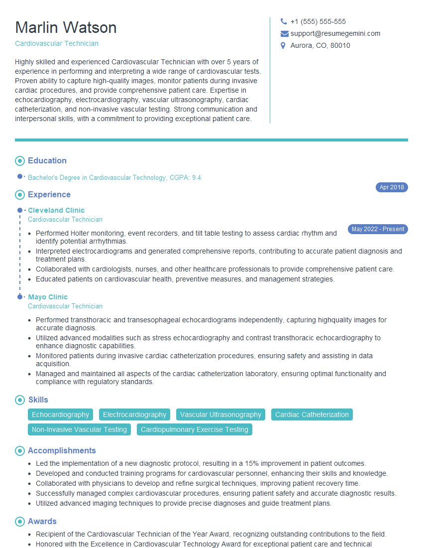
Unlock your full potential by mastering the most common Cardio interview questions. This blog offers a deep dive into the critical topics, ensuring you’re not only prepared to answer but to excel. With these insights, you’ll approach your interview with clarity and confidence.
Questions Asked in Cardio Interview
Q 1. Explain the pathophysiology of coronary artery disease.
Coronary artery disease (CAD) is a condition where plaque builds up inside the coronary arteries, reducing blood flow to the heart muscle. This plaque, composed of cholesterol, fat, calcium, and other substances, is the hallmark of atherosclerosis. The pathophysiology involves several interconnected processes:
- Endothelial Dysfunction: Damage to the inner lining of the arteries initiates the process. This damage can stem from factors like high blood pressure, smoking, diabetes, and high cholesterol.
- Inflammation: Damaged endothelium triggers an inflammatory response, attracting immune cells to the area. These cells contribute to plaque formation.
- Fatty Streak Formation: Low-density lipoprotein (LDL), or “bad” cholesterol, infiltrates the arterial wall, forming fatty streaks – early lesions in atherosclerosis.
- Plaque Development: Over time, these streaks grow, incorporating smooth muscle cells, collagen, and calcium, creating a complex plaque that protrudes into the artery lumen.
- Plaque Rupture and Thrombosis: Plaque can become unstable and rupture, exposing the underlying collagen. This triggers platelet aggregation and blood clot (thrombus) formation, which can completely block blood flow, leading to a heart attack (myocardial infarction).
- Ischemia and Infarction: Reduced blood flow (ischemia) deprives the heart muscle of oxygen and nutrients. If the blockage is severe or prolonged, heart muscle tissue dies (infarction).
Think of it like a pipe gradually clogging with grease; eventually, it can block completely. The severity of CAD depends on the extent and location of the blockage.
Q 2. Describe the different types of cardiomyopathies.
Cardiomyopathies are diseases of the heart muscle itself, weakening its ability to pump blood effectively. They are broadly categorized into:
- Dilated Cardiomyopathy (DCM): The heart chambers become enlarged and weakened, leading to reduced pumping capacity. This can be caused by genetics, infections (like viral myocarditis), alcohol abuse, or chemotherapy.
- Hypertrophic Cardiomyopathy (HCM): The heart muscle thickens, making it harder for the heart to fill with blood. This is often genetic and can lead to sudden cardiac death in young athletes.
- Restrictive Cardiomyopathy (RCM): The heart muscle becomes stiff, limiting its ability to fill with blood. This can be caused by amyloidosis (protein deposits in the heart), radiation therapy, or certain diseases.
- Arrhythmogenic Right Ventricular Cardiomyopathy (ARVC): Fat and fibrous tissue replace heart muscle, particularly in the right ventricle, predisposing to fatal arrhythmias.
Each type has unique symptoms and treatment approaches. For example, DCM might present with shortness of breath and fatigue, while HCM might manifest as chest pain or fainting. Diagnosis involves echocardiography, cardiac MRI, and genetic testing.
Q 3. What are the diagnostic criteria for heart failure?
Diagnosing heart failure involves a combination of clinical evaluation, physical examination, and investigations. Key diagnostic criteria include:
- Symptoms: Shortness of breath (dyspnea), fatigue, ankle swelling (edema), and persistent cough are common.
- Physical Examination: Findings such as abnormal heart sounds (murmurs or gallops), rapid heart rate, and lung crackles support the diagnosis.
- Echocardiography: This ultrasound test assesses heart structure and function, measuring ejection fraction (the percentage of blood pumped out with each beat). Reduced ejection fraction is a hallmark of heart failure, although some patients can have heart failure with preserved ejection fraction (HFpEF).
- Biomarkers: Blood tests measuring levels of natriuretic peptides (BNP or NT-proBNP) help assess the severity of heart failure. Elevated levels are indicative.
- Chest X-ray: Can show signs of fluid buildup in the lungs (pulmonary edema).
The diagnosis is often staged based on the severity of symptoms and the degree of impairment in heart function. This allows for tailored treatment strategies.
Q 4. Discuss the management of acute myocardial infarction.
Management of acute myocardial infarction (AMI), or heart attack, is time-sensitive and focuses on restoring blood flow to the affected area of the heart as quickly as possible. The approach involves:
- Immediate Actions: This includes administering oxygen, aspirin, and nitroglycerin to reduce pain and improve blood flow. If the patient is experiencing cardiac arrest, immediate cardiopulmonary resuscitation (CPR) is vital.
- Reperfusion Therapy: The primary goal is to reopen the blocked coronary artery. This is typically achieved through:
- Percutaneous Coronary Intervention (PCI): A catheter is inserted into the artery, and a balloon is used to inflate the blockage. A stent may be placed to keep the artery open.
- Fibrinolytic Therapy (thrombolysis): Medications are administered intravenously to dissolve the blood clot. This is an alternative to PCI if it is not readily available.
- Ongoing Medical Management: After reperfusion, ongoing treatment includes medications such as beta-blockers, ACE inhibitors, statins, and antiplatelet agents to prevent further complications and reduce long-term risks.
- Cardiac Rehabilitation: A structured program involving exercise, education, and lifestyle modification helps improve heart function and reduce the risk of future events.
Time is muscle in AMI; rapid diagnosis and treatment significantly improve outcomes and reduce mortality.
Q 5. Explain the mechanism of action of statins.
Statins are a class of drugs that effectively lower cholesterol levels in the blood. Their primary mechanism of action is by inhibiting the enzyme HMG-CoA reductase. This enzyme is crucial in the synthesis of cholesterol in the liver.
- HMG-CoA Reductase Inhibition: By blocking this enzyme, statins reduce the liver’s production of cholesterol. This leads to a decrease in LDL (“bad”) cholesterol and an increase in high-density lipoprotein (HDL) (“good”) cholesterol.
- Pleiotropic Effects: Beyond cholesterol lowering, statins have other beneficial effects, such as reducing inflammation, stabilizing plaque, and improving endothelial function. These “pleiotropic effects” contribute to their overall cardiovascular benefits.
The reduction in LDL cholesterol levels achieved by statins translates to a significant decrease in the risk of cardiovascular events, including heart attacks and strokes. It’s important to note that statin therapy is highly effective, but individual responses can vary, and monitoring of lipid levels is important.
Q 6. Describe the various types of cardiac arrhythmias.
Cardiac arrhythmias are abnormalities in the heart’s rhythm. They can range from minor irregularities to life-threatening conditions. Broad categories include:
- Bradyarrhythmias: These are characterized by a slow heart rate. Examples include sinus bradycardia (slow sinus rhythm) and heart blocks (disruptions in the electrical conduction system).
- Tachyarrhythmias: These involve a rapid heart rate. Examples include atrial fibrillation (irregular and rapid atrial rhythm), atrial flutter (rapid atrial rhythm), ventricular tachycardia (rapid ventricular rhythm), and ventricular fibrillation (chaotic and ineffective ventricular rhythm – a life-threatening condition).
- Premature Beats (Extra-systoles): These are extra heartbeats that occur prematurely and can be felt as palpitations.
The specific type of arrhythmia dictates the symptoms and treatment approach. Some arrhythmias may be asymptomatic, while others can cause dizziness, palpitations, chest pain, or even loss of consciousness. Diagnosis involves electrocardiography (ECG) and often cardiac monitoring.
Q 7. What are the indications for cardiac catheterization?
Cardiac catheterization is a minimally invasive procedure used to diagnose and treat various cardiovascular conditions. Indications include:
- Coronary Artery Disease (CAD): To visualize coronary arteries, assess the extent of blockage, and perform PCI if necessary.
- Valvular Heart Disease: To assess the function of heart valves and plan surgical intervention if needed.
- Congenital Heart Defects: To diagnose and assess the severity of congenital heart defects.
- Heart Failure: To assess left ventricular function and pressure measurements.
- Arrhythmias: To perform electrophysiological studies (EPS) to investigate the cause of arrhythmias and potentially ablate (destroy) abnormal electrical pathways.
- Pericardial Disease: To assess pericardial fluid and pressure.
Cardiac catheterization involves inserting a thin, flexible tube (catheter) into a blood vessel, usually in the groin or arm, and guiding it to the heart. The procedure allows for precise visualization of the heart and its blood vessels.
Q 8. Explain the procedure of cardiac pacing.
Cardiac pacing, or pacemaker implantation, is a procedure where a small device is surgically placed under the skin to regulate the heart’s rhythm. It’s used when the heart’s natural electrical system malfunctions, leading to a slow or irregular heartbeat (bradycardia or arrhythmia).
The procedure typically involves making a small incision near the collarbone. A lead wire, which is a thin, insulated wire, is then carefully threaded through a vein to the heart’s chambers. The lead’s tip delivers electrical impulses to stimulate the heart to beat at a normal rate. The other end of the lead connects to the pacemaker generator, a small battery-powered device placed under the skin. This generator monitors the heart’s rhythm and delivers electrical impulses as needed. Post-procedure, patients need to follow specific guidelines to ensure proper healing and functioning of the pacemaker. For example, they might be restricted from heavy lifting for a period.
Think of it like this: your heart is a car engine, and the pacemaker is like a reliable mechanic ensuring the engine starts and runs smoothly, even if it has some electrical issues.
Q 9. Discuss the role of echocardiography in cardiac diagnosis.
Echocardiography, commonly known as an echocardiogram or echo, is a non-invasive imaging technique that uses ultrasound waves to produce real-time images of the heart. It’s a crucial diagnostic tool in cardiology providing detailed information about the heart’s structure and function.
An echo can assess various aspects of cardiac health. It can visualize the heart valves, checking for stenosis (narrowing) or regurgitation (leaking); it can measure the size and thickness of the heart chambers; assess the heart’s pumping ability (ejection fraction); and detect abnormalities such as tumors or blood clots. Different types of echocardiography exist, including transthoracic echocardiography (TTE) which is performed through the chest wall, and transesophageal echocardiography (TEE), which involves placing a small ultrasound probe down the esophagus for a clearer view.
For example, if a patient experiences chest pain and shortness of breath, an echocardiogram can help determine if there is a narrowing of the coronary arteries (indicating coronary artery disease) or if there is a problem with the heart muscle itself. The images and measurements help clinicians to make accurate diagnoses and develop appropriate treatment plans.
Q 10. Describe the different types of heart valves and their functions.
The human heart has four valves that ensure blood flows in one direction only. These valves are crucial for maintaining efficient blood circulation. They open and close rhythmically with each heartbeat, preventing backflow.
- Mitral Valve: Located between the left atrium and left ventricle. It’s a bicuspid valve (two flaps), preventing blood from flowing back into the left atrium when the left ventricle contracts.
- Tricuspid Valve: Located between the right atrium and right ventricle. It’s a tricuspid valve (three flaps) and serves a similar function as the mitral valve, preventing backflow into the right atrium.
- Aortic Valve: Located between the left ventricle and the aorta (the main artery carrying oxygenated blood from the heart to the body). It prevents blood from flowing back into the left ventricle when the heart pumps.
- Pulmonary Valve: Located between the right ventricle and the pulmonary artery (the artery carrying deoxygenated blood to the lungs). It prevents blood from flowing back into the right ventricle.
Valve dysfunction can lead to several conditions like stenosis (narrowing), which restricts blood flow, or regurgitation (leakage), which allows blood to flow backward. These conditions can severely impact the heart’s efficiency and lead to various complications.
Q 11. What are the risk factors for stroke?
Stroke, a serious condition affecting blood supply to the brain, has several modifiable and non-modifiable risk factors.
- Modifiable Risk Factors: These are factors you can control through lifestyle changes or medical treatments. These include: Hypertension (high blood pressure), Atrial fibrillation (irregular heartbeat), High cholesterol, Smoking, Diabetes, Obesity, Physical inactivity, Unhealthy diet.
- Non-Modifiable Risk Factors: These are factors you can’t change. These include: Age (risk increases with age), Family history of stroke, Race (some ethnic groups have a higher risk).
Understanding these risk factors is crucial for prevention and early intervention. For example, maintaining a healthy weight, regular exercise, and a balanced diet can significantly reduce your risk of stroke.
Q 12. Explain the management of hypertension.
Hypertension management aims to lower blood pressure to reduce the risk of cardiovascular complications like stroke and heart attack. Management strategies are typically multi-faceted.
- Lifestyle Modifications: These are crucial first steps and often include weight loss (if overweight or obese), regular aerobic exercise (at least 150 minutes per week), a balanced diet low in sodium and saturated fats, and limiting alcohol intake.
- Medications: If lifestyle modifications are insufficient to control blood pressure, medications are often prescribed. Common drug classes include diuretics (to remove excess fluid), ACE inhibitors or ARBs (to relax blood vessels), beta-blockers (to slow the heart rate), and calcium channel blockers (to relax blood vessels). Medication choice depends on individual factors and other health conditions.
Regular monitoring of blood pressure is crucial to assess the effectiveness of the management plan. Patients should work closely with their healthcare provider to adjust the treatment plan as needed.
Q 13. Discuss the role of lifestyle modifications in cardiovascular health.
Lifestyle modifications play a pivotal role in maintaining cardiovascular health. These changes can significantly reduce your risk of heart disease and stroke.
- Diet: A balanced diet rich in fruits, vegetables, whole grains, and lean proteins is essential. Limiting saturated and trans fats, cholesterol, and sodium is crucial. The Mediterranean diet, for example, has been shown to be beneficial for cardiovascular health.
- Exercise: Regular aerobic exercise, such as brisk walking, swimming, or cycling, strengthens the heart and improves blood circulation. Aim for at least 150 minutes of moderate-intensity aerobic activity or 75 minutes of vigorous-intensity aerobic activity per week.
- Weight Management: Maintaining a healthy weight reduces the strain on the heart and blood vessels.
- Stress Management: Chronic stress can negatively affect cardiovascular health. Stress-reducing techniques like yoga, meditation, or deep breathing can be beneficial.
- Smoking Cessation: Smoking is a major risk factor for cardiovascular disease. Quitting smoking significantly reduces your risk.
- Alcohol Consumption: Moderate alcohol consumption may have some benefits, but excessive drinking increases the risk of cardiovascular diseases.
These lifestyle changes are not only preventive measures but also contribute to overall well-being and improved quality of life. Even small changes can make a big difference in the long run.
Q 14. What are the common complications of cardiac surgery?
Cardiac surgery, while life-saving, carries potential complications. These can range from minor to life-threatening.
- Bleeding: Excessive bleeding at the surgical site is a potential risk, requiring close monitoring and sometimes blood transfusions.
- Infection: Infections at the incision site or within the heart are possible and can be serious, requiring treatment with antibiotics.
- Arrhythmias: Irregular heartbeats can occur as a result of the surgery, sometimes requiring medication or further procedures.
- Stroke: Blood clots can form during or after surgery, potentially traveling to the brain and causing a stroke.
- Kidney Failure: The use of certain medications or the effects of the surgery itself can sometimes lead to kidney problems.
- Heart Failure: In some cases, the surgery might not fully resolve the underlying heart issue, and heart failure can occur or worsen.
The risk of these complications varies depending on factors such as the patient’s overall health, the type of surgery performed, and the skill of the surgical team. Post-operative care is crucial in minimizing these risks and ensuring successful recovery.
Q 15. Explain the principles of cardiac rehabilitation.
Cardiac rehabilitation is a structured program designed to help individuals recover from cardiovascular events like heart attacks, heart surgery, or heart failure. It focuses on improving cardiovascular health, increasing physical fitness, and promoting healthy lifestyle changes. The core principles are multidisciplinary, encompassing medical, psychological, and lifestyle interventions.
- Graded Exercise: This involves a carefully planned exercise program that gradually increases in intensity and duration, tailored to the individual’s capabilities and monitored by healthcare professionals. Think of it like gently retraining your heart muscle to work more efficiently.
- Risk Factor Modification: This addresses lifestyle factors that contribute to heart disease. This includes smoking cessation, dietary changes (like lowering sodium and saturated fat intake), weight management, stress reduction techniques, and medication adherence.
- Education and Counseling: Patients receive detailed education on their condition, medication management, and strategies to prevent future events. Psychological support is crucial to address anxiety, depression, and other emotional challenges that can arise after a cardiac event.
- Self-Management Strategies: The goal is to empower patients to take control of their own health. This includes teaching patients how to monitor their symptoms, recognize warning signs, and manage their medications effectively. Patients are encouraged to become active participants in their recovery journey.
For example, a patient recovering from a heart attack might start with short walks, gradually increasing the distance and intensity over several weeks, guided by their healthcare team. They might also receive dietary counseling to help them adopt a heart-healthy eating plan and participate in stress management workshops.
Career Expert Tips:
- Ace those interviews! Prepare effectively by reviewing the Top 50 Most Common Interview Questions on ResumeGemini.
- Navigate your job search with confidence! Explore a wide range of Career Tips on ResumeGemini. Learn about common challenges and recommendations to overcome them.
- Craft the perfect resume! Master the Art of Resume Writing with ResumeGemini’s guide. Showcase your unique qualifications and achievements effectively.
- Don’t miss out on holiday savings! Build your dream resume with ResumeGemini’s ATS optimized templates.
Q 16. How do you interpret an electrocardiogram (ECG)?
Interpreting an electrocardiogram (ECG) involves analyzing the electrical activity of the heart. It’s like reading the heart’s electrical roadmap. The ECG tracing displays waveforms representing the depolarization (electrical activation) and repolarization (electrical recovery) of the atria and ventricles.
- P wave: Represents atrial depolarization.
- QRS complex: Represents ventricular depolarization.
- T wave: Represents ventricular repolarization.
Interpretation involves assessing the following:
- Rhythm: Is the heart beating regularly or irregularly? Are there any extra beats or pauses?
- Rate: What is the heart rate (number of beats per minute)?
- Axis: What is the overall direction of electrical activity in the heart?
- Intervals and segments: Analyzing the durations of different segments (e.g., PR interval, QT interval) can identify conduction delays or abnormalities.
- Waveforms: The shape and amplitude of the waves can indicate abnormalities such as hypertrophy (enlarged heart muscle), ischemia (reduced blood flow), or infarction (heart attack).
For instance, a prolonged QRS complex might suggest a bundle branch block, a condition where the electrical signal doesn’t travel properly through the ventricles. A ST-segment elevation might indicate a heart attack requiring immediate intervention. ECG interpretation requires training and experience, and any significant abnormalities should be promptly reviewed by a cardiologist.
Q 17. Describe the different types of cardiac imaging modalities.
Cardiac imaging modalities are non-invasive and invasive techniques used to visualize the heart and assess its structure and function. They provide detailed information about the heart’s chambers, valves, blood vessels, and electrical activity.
- Echocardiography: Uses ultrasound waves to create images of the heart. It’s the most commonly used cardiac imaging technique and is excellent for assessing valve function, wall motion, and chamber size. Think of it like an ultrasound for your heart. There are different types of echocardiograms, including transthoracic (TTE) and transesophageal (TEE).
- Cardiac Computed Tomography (CT): Uses X-rays to generate detailed cross-sectional images of the heart. It’s particularly useful for assessing coronary artery disease and identifying calcium deposits in the arteries.
- Cardiac Magnetic Resonance Imaging (MRI): Uses a powerful magnetic field and radio waves to produce images of the heart. It’s useful for evaluating heart muscle function, assessing the extent of damage from a heart attack, and detecting congenital heart defects.
- Nuclear Cardiology: Uses radioactive tracers to assess blood flow to the heart muscle. It is useful for detecting coronary artery disease and evaluating the effectiveness of revascularization procedures.
- Coronary Angiography: An invasive procedure where a catheter is inserted into a blood vessel and guided to the heart to visualize the coronary arteries. It is the gold standard for diagnosing coronary artery disease and performing interventional procedures like angioplasty and stenting.
The choice of imaging modality depends on the specific clinical question and the patient’s condition. For example, an echocardiogram is often the first-line investigation for suspected valvular heart disease, while coronary angiography is indicated when coronary artery disease is suspected.
Q 18. What are the contraindications for cardiac procedures?
Contraindications for cardiac procedures refer to situations where the risks of the procedure outweigh the potential benefits. These can be broadly categorized into relative and absolute contraindications. Relative contraindications mean the procedure might be considered if the benefits outweigh the risks, while absolute contraindications generally preclude the procedure.
- Uncontrolled bleeding disorders: This increases the risk of hemorrhage during and after the procedure.
- Severe renal or hepatic impairment: This affects the body’s ability to metabolize contrast agents and medications used during the procedure.
- Uncontrolled hypertension (high blood pressure): This poses an increased risk of complications during the procedure.
- Severe infections: Increased risk of infection and poor wound healing.
- Severe allergic reactions to contrast media: Contrast agents are used in many cardiac procedures (e.g., angiography), and severe allergic reactions can be life-threatening.
- Active endocarditis (infection of the heart lining): This increases the risk of spreading the infection.
- Recent myocardial infarction (heart attack): The heart might not be stable enough to tolerate the procedure.
Each patient’s situation is unique and the decision to proceed with a cardiac procedure requires careful consideration of the individual’s medical history, current condition, and the potential benefits and risks of the procedure. For example, a patient with severe, uncontrolled bleeding might be deemed ineligible for a coronary artery bypass graft (CABG) due to the high risk of bleeding complications. A thorough evaluation by a cardiologist and the cardiology team is essential before any cardiac procedure.
Q 19. Explain the use of anticoagulants in cardiovascular disease.
Anticoagulants, also known as blood thinners, are medications used to prevent blood clots. They play a vital role in the management and prevention of cardiovascular disease by reducing the risk of thromboembolic events (blood clots that travel to and block blood vessels).
- Atrial fibrillation: Anticoagulants are crucial in reducing the risk of stroke in patients with atrial fibrillation, a common heart rhythm disorder that increases the risk of clot formation in the heart.
- Deep vein thrombosis (DVT) and pulmonary embolism (PE): Anticoagulants are used to treat and prevent DVT (blood clots in the deep veins of the legs) and PE (blood clots that travel to the lungs).
- After cardiac procedures: Anticoagulants are often prescribed after procedures like heart valve replacement or coronary artery bypass grafting to prevent clot formation.
- Myocardial infarction (heart attack): Anticoagulants might be used to prevent further clot formation in the coronary arteries after a heart attack.
Different types of anticoagulants exist, including warfarin, heparin, direct thrombin inhibitors (e.g., dabigatran), and direct factor Xa inhibitors (e.g., rivaroxaban). The choice of anticoagulant depends on various factors, including the patient’s specific condition, other medical issues, and potential drug interactions. Regular monitoring is necessary to ensure the effectiveness and safety of anticoagulant therapy. For example, patients on warfarin require regular blood tests to monitor their INR (international normalized ratio) to ensure the medication is working effectively and to avoid excessive bleeding.
Q 20. Discuss the role of genetic factors in cardiovascular disease.
Genetic factors play a significant role in the development of cardiovascular disease (CVD). While lifestyle factors are also crucial, inherited traits can increase susceptibility to various heart conditions.
- Familial Hypercholesterolemia: An inherited disorder characterized by high levels of cholesterol in the blood. This greatly increases the risk of early atherosclerosis (hardening of the arteries) and coronary artery disease.
- Polygenic Risk Scores: These scores combine information from multiple genetic variations to estimate an individual’s risk of developing CVD. The higher the score, the greater the risk.
- Specific Gene Mutations: Certain gene mutations are associated with an increased risk of specific cardiac conditions. For instance, mutations in genes related to the production of collagen can increase the risk of aortic aneurysms (bulging of the aorta).
- Inherited Arrhythmias: Genetic factors can increase susceptibility to certain heart rhythm disorders, like Long QT syndrome, which can lead to potentially fatal arrhythmias.
Understanding the role of genetics in CVD is becoming increasingly important. Genetic testing can help identify individuals at increased risk, allowing for preventive strategies and early intervention. For example, a family history of early heart disease warrants a thorough cardiovascular risk assessment and lifestyle modifications to mitigate potential risks. This underlines the importance of family history in CVD risk assessment.
Q 21. How do you assess the risk of cardiovascular events?
Assessing the risk of cardiovascular events involves evaluating various risk factors to predict the likelihood of developing or experiencing a heart attack, stroke, or other cardiovascular complications. This often involves a combination of clinical assessment, laboratory tests, and risk scoring systems.
- Risk Factors: These include age, sex, family history of CVD, smoking, hypertension, high cholesterol, diabetes, obesity, physical inactivity, and unhealthy diet.
- Clinical Assessment: This involves taking a thorough medical history, performing a physical examination, and reviewing the patient’s medications.
- Laboratory Tests: Laboratory tests such as lipid profiles (cholesterol levels), blood glucose, and kidney function tests provide additional information.
- Risk Scoring Systems: Standardized scoring systems, such as the Framingham Risk Score or the Reynolds Risk Score, use risk factors to calculate an individual’s 10-year risk of developing CVD. These scores help guide treatment decisions and preventive strategies.
- Imaging Studies: Cardiac imaging (e.g., echocardiography, coronary angiography) may be used to assess the presence and extent of underlying heart disease.
For example, a 60-year-old male smoker with high cholesterol, hypertension, and a family history of early heart disease would be considered at high risk of a cardiovascular event. This individual would likely benefit from aggressive risk factor modification, including lifestyle changes and medication. The goal of risk assessment is to identify individuals at higher risk, enabling proactive interventions to prevent or delay cardiovascular events and improve patient outcomes. A multifactorial approach considering various factors is key to an accurate risk stratification.
Q 22. Describe the different types of cardiac pacemakers.
Cardiac pacemakers are life-saving devices that help regulate the heart’s rhythm. They come in various types, primarily categorized by their function and placement.
- Single-Chamber Pacemakers: These pace only one chamber of the heart, either the right atrium (A) or the right ventricle (V). They’re suitable for patients with slow heartbeats originating in the atria or ventricles.
- Dual-Chamber Pacemakers (AAI, VVI, DDD): These pace both the atria and ventricles, mimicking the natural heart rhythm more closely. ‘DDD’ pacemakers can sense and respond to both atrial and ventricular activity, offering the most comprehensive pacing.
- Biventricular Pacemakers (CRT-P or CRT-D): These are used for patients with heart failure and conduction delays. They deliver electrical impulses to both ventricles, improving heart function and reducing symptoms. The ‘D’ signifies that it also includes defibrillation capabilities.
- Leadless Pacemakers: These are small, self-contained pacemakers implanted directly into the heart’s right ventricle without the need for leads. They are minimally invasive and offer improved longevity in some cases.
The choice of pacemaker depends on the patient’s specific condition and needs. For instance, a patient with atrial fibrillation might benefit from a DDD pacemaker, while a patient with a slow ventricular rhythm might only require a VVI pacemaker.
Q 23. Explain the principles of hemodynamic monitoring.
Hemodynamic monitoring measures the heart’s ability to pump blood and the circulatory system’s response. It involves assessing various parameters to understand the cardiovascular system’s overall performance.
- Blood Pressure: This measures the force of blood against artery walls, reflecting the heart’s pumping strength (systolic) and the artery’s resistance (diastolic). Sustained hypertension or hypotension indicates serious cardiovascular issues.
- Heart Rate: The number of times the heart beats per minute provides insight into its rhythm and response to stress or illness. Tachycardia (high heart rate) and bradycardia (low heart rate) need evaluation.
- Cardiac Output: This measures the volume of blood pumped by the heart per minute, indicating its overall efficiency. It reflects stroke volume (amount of blood pumped per beat) and heart rate.
- Central Venous Pressure (CVP): This reflects the pressure in the vena cava, providing information about fluid status and right heart function. Elevated CVP suggests fluid overload.
- Pulmonary Artery Pressure (PAP): This assesses pressure in the pulmonary artery, crucial for evaluating pulmonary circulation and the left heart’s function. High PAP could suggest pulmonary hypertension.
Monitoring these parameters helps clinicians identify and manage conditions like heart failure, shock, and other cardiovascular complications. For example, a patient in cardiogenic shock might exhibit low blood pressure, high heart rate, and low cardiac output, prompting immediate intervention.
Q 24. What are the different types of heart transplants?
While the term ‘different types of heart transplants’ isn’t strictly accurate – a heart transplant is a heart transplant – we can categorize them based on the source of the donor heart and the recipient’s condition:
- Orthotopic Heart Transplantation: This is the most common type, where the diseased heart is removed and replaced with a healthy donor heart. The donor heart is positioned in the same anatomical location as the original heart.
- Heterotopic Heart Transplantation: In this less common procedure, the donor heart is implanted alongside the patient’s diseased heart. This is generally reserved for patients with irreversible heart failure but still with some residual function in their native heart.
The choice between these approaches depends on the severity of the recipient’s condition and the suitability of the donor heart. The vast majority of heart transplants are orthotopic.
Q 25. Describe the role of implantable cardioverter-defibrillators (ICDs).
Implantable cardioverter-defibrillators (ICDs) are sophisticated devices that prevent sudden cardiac death in patients at high risk. They monitor the heart’s rhythm and deliver therapies when necessary.
- Detection of Lethal Rhythms: ICDs continuously monitor the heart’s rhythm for life-threatening arrhythmias like ventricular tachycardia (VT) or ventricular fibrillation (VF).
- Cardioversion: If the ICD detects a rapid but potentially reversible arrhythmia (VT), it delivers a synchronized electrical shock (cardioversion) to restore normal rhythm.
- Defibrillation: If the ICD detects a life-threatening arrhythmia (VF), it delivers an unsynchronized high-energy shock (defibrillation) to stop the chaotic electrical activity and allow the heart to resume a normal rhythm.
- Anti-tachycardia Pacing (ATP): Some ICDs can also deliver rapid pacing impulses to terminate some tachycardias before they escalate to life-threatening arrhythmias.
ICDs are essential for patients who have survived a cardiac arrest, have a history of life-threatening arrhythmias, or have heart conditions predisposing them to sudden death. For example, a patient with a history of ventricular fibrillation would greatly benefit from an ICD to prevent recurrence.
Q 26. Explain the management of hyperlipidemia.
Hyperlipidemia, or high cholesterol, is managed through a multifaceted approach focusing on lifestyle modifications and medication when necessary.
- Dietary Changes: A diet low in saturated and trans fats, cholesterol, and sodium, while rich in fruits, vegetables, and fiber, is crucial. This helps lower LDL (‘bad’) cholesterol and raise HDL (‘good’) cholesterol.
- Exercise: Regular physical activity helps raise HDL cholesterol and lower LDL cholesterol and triglycerides. Aim for at least 150 minutes of moderate-intensity or 75 minutes of vigorous-intensity aerobic exercise per week.
- Weight Management: Losing even a small amount of weight can significantly improve lipid profiles.
- Smoking Cessation: Smoking elevates LDL cholesterol and lowers HDL cholesterol, significantly increasing cardiovascular risk. Cessation is vital.
- Medications: Statins are the first-line treatment for most patients with hyperlipidemia. They effectively lower LDL cholesterol. Other medications, such as fibrates or ezetimibe, may be added if needed.
For example, a patient with high LDL cholesterol might start with dietary changes and exercise. If those measures aren’t sufficient, a statin medication would be introduced. Regular monitoring of cholesterol levels is essential to assess treatment effectiveness.
Q 27. What is your experience with different types of cardiac devices?
Throughout my career, I’ve extensively worked with various cardiac devices, including pacemakers, ICDs, and cardiac resynchronization therapy (CRT) devices. My experience encompasses implantation, programming, troubleshooting, and follow-up care. I’m proficient in interpreting device data, identifying malfunctions, and managing complications. I’ve also been involved in research evaluating the efficacy and safety of new cardiac device technologies.
For instance, I’ve managed patients with lead failures in pacemakers, requiring prompt intervention and lead extraction or replacement. I’ve also worked with patients experiencing inappropriate shocks from ICDs, necessitating programming adjustments or device upgrades. This extensive experience has given me a solid understanding of the nuances of each device and the associated complexities in patient management.
Q 28. How do you handle a patient with a cardiac emergency?
Managing a cardiac emergency requires immediate, decisive action. The approach follows a structured protocol prioritizing airway, breathing, and circulation (ABCs).
- Assess the Situation: Quickly evaluate the patient’s condition, identifying the nature of the cardiac emergency (e.g., cardiac arrest, acute coronary syndrome).
- Activate Emergency Response: Immediately call for emergency medical services (EMS) and initiate advanced cardiac life support (ACLS) if qualified.
- Airway, Breathing, Circulation (ABCs): Ensure a patent airway, provide rescue breaths if necessary, and initiate cardiopulmonary resuscitation (CPR) if the patient is pulseless and breathless.
- Defibrillation (if applicable): If a defibrillator is available and the patient is in ventricular fibrillation or pulseless ventricular tachycardia, deliver immediate defibrillation.
- Medication Administration (if applicable): Administer appropriate medications according to ACLS guidelines (e.g., epinephrine, amiodarone).
- Continuous Monitoring: Continuously monitor vital signs, ECG rhythm, and response to treatment.
- Post-Resuscitation Care: Following resuscitation, provide supportive care including oxygen, IV fluids, and ongoing ECG monitoring. Transfer the patient to a hospital for definitive care.
For example, if a patient collapses and becomes unresponsive, immediate CPR and defibrillation (if available) are crucial. Simultaneously, EMS should be contacted to provide advanced life support and transport to the hospital.
Key Topics to Learn for Your Cardio Interview
- Cardiac Physiology: Understand the intricacies of the heart’s electrical conduction system, contractile properties, and hemodynamics. Consider exploring the Frank-Starling mechanism and its clinical relevance.
- Cardiac Pathology: Develop a strong understanding of common cardiac conditions such as coronary artery disease, heart failure, arrhythmias (e.g., atrial fibrillation, ventricular tachycardia), valvular heart disease, and congenital heart defects. Focus on pathophysiological mechanisms and clinical presentations.
- Diagnostic Techniques: Familiarize yourself with the interpretation of electrocardiograms (ECGs), echocardiograms, cardiac catheterization results, and stress tests. Understand the strengths and limitations of each technique.
- Pharmacology: Gain a solid understanding of the mechanisms of action, indications, contraindications, and side effects of common cardiovascular medications, including antiarrhythmics, antihypertensives, anticoagulants, and statins.
- Treatment Strategies: Explore various treatment approaches for cardiovascular diseases, encompassing medical management, surgical interventions (e.g., coronary artery bypass grafting, valve replacement), and interventional cardiology procedures (e.g., angioplasty, stent placement).
- Clinical Reasoning and Problem Solving: Practice applying your knowledge to solve clinical case studies. Focus on identifying key symptoms, interpreting diagnostic findings, and formulating appropriate management plans.
- Research and Current Trends: Stay updated on recent advancements in cardiovascular research and emerging treatment strategies. Demonstrating awareness of cutting-edge techniques will impress interviewers.
Next Steps
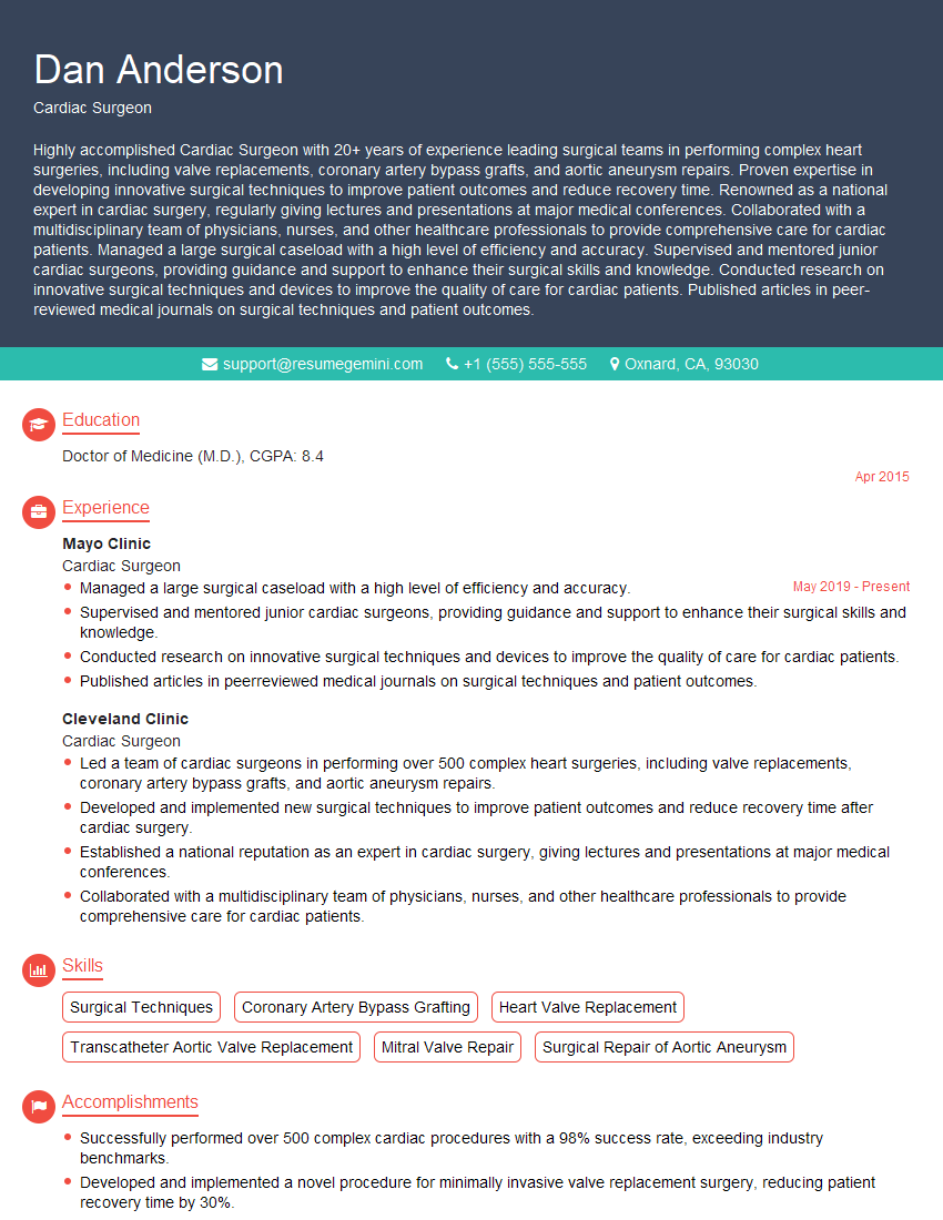
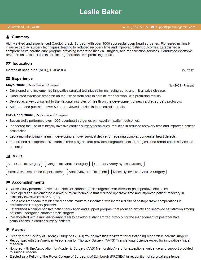
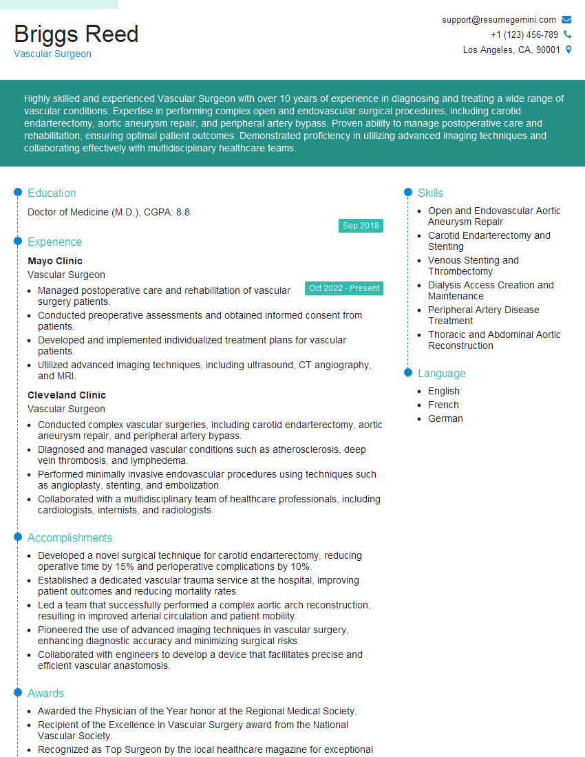
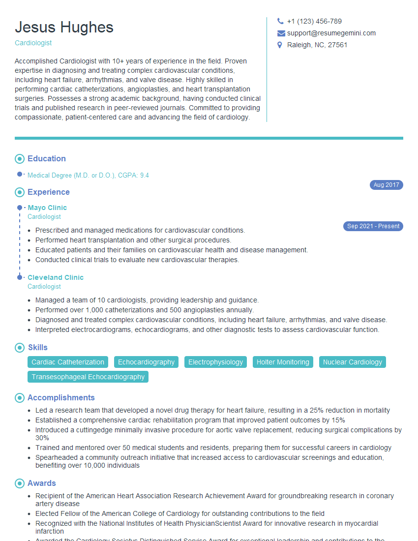
Mastering these key areas in cardiology will significantly enhance your career prospects, opening doors to exciting opportunities in research, clinical practice, or industry. To maximize your chances of success, it’s crucial to present your skills and experience effectively. Creating a strong, ATS-friendly resume is paramount. ResumeGemini is a trusted resource that can help you build a professional and impactful resume, tailored to highlight your unique qualifications within the competitive field of cardiology. Examples of resumes specifically designed for cardiologist positions are available to guide you.
Explore more articles
Users Rating of Our Blogs
Share Your Experience
We value your feedback! Please rate our content and share your thoughts (optional).
What Readers Say About Our Blog
Hello,
We found issues with your domain’s email setup that may be sending your messages to spam or blocking them completely. InboxShield Mini shows you how to fix it in minutes — no tech skills required.
Scan your domain now for details: https://inboxshield-mini.com/
— Adam @ InboxShield Mini
Reply STOP to unsubscribe
Hi, are you owner of interviewgemini.com? What if I told you I could help you find extra time in your schedule, reconnect with leads you didn’t even realize you missed, and bring in more “I want to work with you” conversations, without increasing your ad spend or hiring a full-time employee?
All with a flexible, budget-friendly service that could easily pay for itself. Sounds good?
Would it be nice to jump on a quick 10-minute call so I can show you exactly how we make this work?
Best,
Hapei
Marketing Director
Hey, I know you’re the owner of interviewgemini.com. I’ll be quick.
Fundraising for your business is tough and time-consuming. We make it easier by guaranteeing two private investor meetings each month, for six months. No demos, no pitch events – just direct introductions to active investors matched to your startup.
If youR17;re raising, this could help you build real momentum. Want me to send more info?
Hi, I represent an SEO company that specialises in getting you AI citations and higher rankings on Google. I’d like to offer you a 100% free SEO audit for your website. Would you be interested?
Hi, I represent an SEO company that specialises in getting you AI citations and higher rankings on Google. I’d like to offer you a 100% free SEO audit for your website. Would you be interested?
good
