Preparation is the key to success in any interview. In this post, we’ll explore crucial Knowledge of medical terminology and anatomy interview questions and equip you with strategies to craft impactful answers. Whether you’re a beginner or a pro, these tips will elevate your preparation.
Questions Asked in Knowledge of medical terminology and anatomy Interview
Q 1. Define ‘osteoporosis’ and explain its impact on bone structure.
Osteoporosis is a systemic skeletal disease characterized by low bone mass and microarchitectural deterioration of bone tissue, leading to increased bone fragility and a consequent increase in fracture risk. Imagine your bones as a honeycomb – in osteoporosis, the honeycomb structure becomes thinner and weaker, making it more susceptible to breaks. This happens because of an imbalance between bone resorption (breakdown) and bone formation (building). The body constantly remodels bone, but in osteoporosis, the resorption process outpaces bone formation, resulting in a net loss of bone mass.
The impact on bone structure is significant. Bones become porous and less dense, making them more fragile and prone to fractures, even from minor falls or trauma. These fractures can occur in various locations, including the hip, spine, and wrist, leading to significant pain, disability, and reduced quality of life. Common complications include vertebral compression fractures leading to loss of height and kyphosis (curvature of the spine), as well as hip fractures which can result in prolonged hospitalization and loss of mobility.
Q 2. What is the difference between ‘proximal’ and ‘distal’?
Proximal and distal are terms used to describe the relative position of a body part along a limb or appendage. Think of your arm: your shoulder is proximal to your elbow, and your elbow is proximal to your wrist. Your wrist is distal to your elbow, and your hand is distal to your wrist.
In simpler terms, proximal means closer to the point of attachment (like the torso) and distal means further away from the point of attachment. This terminology is crucial in anatomical descriptions and medical documentation to precisely locate structures and injuries.
Q 3. Explain the function of the cerebellum.
The cerebellum, located at the back of the brain, is primarily responsible for coordinating voluntary movements, maintaining balance and posture, and motor learning. Think of it as the brain’s ‘fine-tuning’ center for movement. It receives input from various parts of the brain and the sensory systems to refine motor commands, ensuring smooth, coordinated actions.
Its functions go beyond just movement; the cerebellum also plays a subtle role in cognitive functions like language and attention. Damage to the cerebellum can lead to ataxia (lack of coordination), tremors, difficulty with balance, and problems with motor learning. For example, a person with cerebellar damage might struggle to walk in a straight line or perform simple tasks requiring precise hand-eye coordination.
Q 4. What is the medical term for inflammation of the liver?
The medical term for inflammation of the liver is hepatitis. Hepatitis is not a single disease but rather a group of diseases that cause liver inflammation. Various factors can cause hepatitis, including viral infections (hepatitis A, B, C, D, and E), autoimmune disorders, alcohol abuse, and certain medications. The symptoms can range from mild to severe and may include jaundice (yellowing of the skin and eyes), abdominal pain, fatigue, and nausea.
Q 5. Define ‘dysplasia’ and give an example.
Dysplasia refers to the abnormal development or growth of cells, tissues, or organs. It’s a precancerous condition, meaning it is an abnormal change that may progress to cancer but does not always do so. Instead of normal, orderly cellular growth, dysplasia shows disorganized cellular arrangement, altered cell size and shape, and an increased number of dividing cells.
A common example is cervical dysplasia, where abnormal cells develop on the cervix. This is often detected through Pap smears and is usually treatable if caught early. However, if left untreated, it can potentially develop into cervical cancer. Dysplasia can occur in various organs and tissues throughout the body.
Q 6. What is the difference between ‘benign’ and ‘malignant’ tumors?
The key difference between benign and malignant tumors lies in their behavior and potential to spread. Benign tumors are generally slow-growing, localized, and do not invade surrounding tissues or metastasize (spread to other parts of the body). They are typically encapsulated, meaning they have a defined border, and are usually non-life-threatening unless they compress vital organs.
Malignant tumors, on the other hand, are cancerous. They grow rapidly, invade surrounding tissues, and can metastasize through the bloodstream or lymphatic system to distant sites in the body. This metastatic spread is what makes malignant tumors life-threatening. They are not encapsulated and can be difficult to remove completely.
Q 7. Explain the process of cellular respiration.
Cellular respiration is the process by which cells break down glucose (a sugar) to generate energy in the form of ATP (adenosine triphosphate). It’s a series of metabolic reactions that occur within the mitochondria (the powerhouses of the cell). Think of it as the cell’s way of ‘burning’ fuel to produce energy for its functions.
The process can be broadly divided into three main stages:
- Glycolysis: Glucose is broken down in the cytoplasm into pyruvate.
- Krebs Cycle (Citric Acid Cycle): Pyruvate is further oxidized in the mitochondria, releasing carbon dioxide and generating ATP and electron carriers.
- Electron Transport Chain: The electron carriers transfer electrons through a series of protein complexes embedded in the mitochondrial membrane. This process generates a proton gradient, which drives ATP synthesis.
The ATP produced powers various cellular activities, including muscle contraction, protein synthesis, and maintaining cell membrane integrity. Cellular respiration is essential for life as it provides the energy needed for all cellular processes.
Q 8. What is the function of the endocrine system?
The endocrine system is a complex network of glands that produce and secrete hormones directly into the bloodstream. These hormones act as chemical messengers, traveling throughout the body to regulate a vast array of physiological processes. Think of it as the body’s internal communication system, using hormones instead of nerves.
Its functions include regulating metabolism, growth and development, tissue function, sexual function, reproduction, sleep, and mood, among many others. For example, the thyroid gland produces hormones that control metabolism; the pancreas releases insulin to regulate blood sugar; and the adrenal glands release adrenaline in response to stress. Dysfunction in any part of the endocrine system can lead to a wide range of disorders, highlighting its crucial role in maintaining overall health.
Q 9. Describe the structure and function of the nephron.
The nephron is the functional unit of the kidney, responsible for filtering blood and producing urine. Imagine it as a tiny, intricate filter within the kidney. Each kidney contains millions of nephrons.
Its structure involves several key components:
- Renal corpuscle: This includes the glomerulus (a network of capillaries) and Bowman’s capsule (a cup-like structure surrounding the glomerulus). Blood is initially filtered here.
- Renal tubule: This long, twisted tube consists of the proximal convoluted tubule, loop of Henle, and distal convoluted tubule. As the filtrate passes through these sections, essential substances like water, glucose, and amino acids are reabsorbed back into the bloodstream, while waste products are concentrated.
- Collecting duct: Multiple nephrons share a collecting duct, which carries the final urine to the renal pelvis and eventually to the ureter.
The function is essentially a three-step process: filtration, reabsorption, and secretion. Filtration occurs in the glomerulus, removing water and small solutes from the blood. Reabsorption reclaims essential substances. Secretion actively removes additional wastes and excess ions. The ultimate product is urine, which carries waste products out of the body.
Q 10. What are the main components of blood?
Blood is a specialized connective tissue with several vital components:
- Plasma: This is the liquid part of the blood, comprising about 55% of its volume. It consists mainly of water, proteins (like albumin, globulins, and fibrinogen), electrolytes, nutrients, hormones, and waste products.
- Red blood cells (Erythrocytes): These are the most abundant cells in blood and contain hemoglobin, a protein that carries oxygen from the lungs to the body’s tissues and carbon dioxide back to the lungs. Their primary function is gas transport.
- White blood cells (Leukocytes): These cells are part of the immune system, defending the body against infection and disease. Several types exist, each with a specific role.
- Platelets (Thrombocytes): These cell fragments are crucial for blood clotting (coagulation), preventing excessive bleeding from injuries.
The balance and proper function of these components are critical for maintaining overall health. For example, a deficiency in red blood cells can lead to anemia, while a low platelet count can cause excessive bleeding.
Q 11. Explain the difference between arteries and veins.
Arteries and veins are both types of blood vessels, but they have distinct differences in structure and function:
- Arteries: Carry oxygenated blood away from the heart to the body’s tissues (except for pulmonary arteries, which carry deoxygenated blood to the lungs). They have thick, elastic walls to withstand the high pressure of blood pumped from the heart. Their walls contain smooth muscle that can constrict or dilate to regulate blood flow.
- Veins: Carry deoxygenated blood back to the heart from the tissues (except for pulmonary veins, which carry oxygenated blood from the lungs to the heart). They have thinner walls than arteries and contain valves to prevent backflow of blood, as the pressure is lower.
Think of arteries as high-pressure, high-speed highways carrying oxygen-rich blood, while veins are like lower-pressure, slower-moving roads bringing deoxygenated blood back to the heart for re-oxygenation. These differences in structure reflect their distinct roles in the circulatory system.
Q 12. What is the role of the lymphatic system?
The lymphatic system is a network of tissues, vessels, and organs that work together to maintain fluid balance, absorb fats, and defend the body against infection. It plays a crucial supporting role to the circulatory system and immune system.
Its main functions include:
- Fluid balance: It collects excess fluid (lymph) from tissues and returns it to the bloodstream, preventing fluid buildup.
- Fat absorption: It absorbs fats and fat-soluble vitamins from the digestive system and transports them to the bloodstream.
- Immune defense: It contains lymphocytes (a type of white blood cell) and other immune cells that help fight infections and diseases. Lymph nodes, which are located throughout the lymphatic system, act as filters, trapping pathogens and other harmful substances.
Imagine the lymphatic system as a drainage and defense system for the body, removing excess fluid and fighting off invaders. Swelling (edema) can occur if the lymphatic system is not functioning properly.
Q 13. Define ‘aphasia’ and its different types.
Aphasia is a language disorder affecting the ability to communicate. It’s caused by damage to the parts of the brain that control language. This damage can stem from stroke, brain injury, or neurological conditions.
Different types of aphasia exist, impacting different aspects of language:
- Broca’s aphasia (expressive aphasia): Difficulty producing speech, often characterized by short, fragmented sentences. Understanding is typically better than speaking.
- Wernicke’s aphasia (receptive aphasia): Difficulty understanding spoken or written language, even though speech may be fluent but nonsensical (“word salad”).
- Global aphasia: Severe impairment in both comprehension and production of speech.
- Conduction aphasia: Difficulty repeating words or phrases, though comprehension and spontaneous speech are relatively preserved.
- Anomic aphasia: Difficulty retrieving words, often leading to word-finding pauses and substitutions.
The specific type of aphasia and its severity depend on the location and extent of brain damage. Rehabilitation is crucial to improve communication skills.
Q 14. What is the difference between ‘tachycardia’ and ‘bradycardia’?
Both tachycardia and bradycardia describe abnormalities in heart rate, but in opposite directions:
- Tachycardia: A heart rate that is abnormally fast, typically exceeding 100 beats per minute (bpm) at rest. This can be caused by various factors including exercise, stress, fever, dehydration, and heart conditions.
- Bradycardia: A heart rate that is abnormally slow, typically below 60 bpm at rest. This can be due to factors like medication, electrolyte imbalances, heart conditions, or sleep.
While a slightly elevated or lowered heart rate might be normal in certain circumstances, consistently fast (tachycardia) or slow (bradycardia) heart rates can be indicative of underlying health issues and require medical attention. They can both potentially impact the body’s ability to deliver sufficient oxygen to tissues.
Q 15. Explain the function of the pancreas.
The pancreas is a fascinating organ with both endocrine and exocrine functions. Think of it as a dual-purpose gland.
Exocrine function: This involves the production and secretion of digestive enzymes. These enzymes, including amylase (for carbohydrate digestion), lipase (for fat digestion), and protease (for protein digestion), are released into the duodenum (the first part of the small intestine) via the pancreatic duct. These enzymes are crucial for breaking down food into smaller molecules that can be absorbed into the bloodstream. Imagine them as tiny demolition crews breaking down the food ‘bricks’ into smaller, manageable pieces.
Endocrine function: This involves the production and secretion of hormones directly into the bloodstream. The most important hormones produced by the pancreas are insulin and glucagon. Insulin helps regulate blood sugar levels by allowing glucose to enter cells, while glucagon raises blood sugar levels when they fall too low. This is like a finely tuned thermostat regulating blood sugar levels, preventing both highs (hyperglycemia) and lows (hypoglycemia).
In essence, the pancreas plays a vital role in both digestion and blood sugar regulation, highlighting its importance for overall health.
Career Expert Tips:
- Ace those interviews! Prepare effectively by reviewing the Top 50 Most Common Interview Questions on ResumeGemini.
- Navigate your job search with confidence! Explore a wide range of Career Tips on ResumeGemini. Learn about common challenges and recommendations to overcome them.
- Craft the perfect resume! Master the Art of Resume Writing with ResumeGemini’s guide. Showcase your unique qualifications and achievements effectively.
- Don’t miss out on holiday savings! Build your dream resume with ResumeGemini’s ATS optimized templates.
Q 16. What are the symptoms of appendicitis?
Appendicitis is inflammation of the appendix, a small, finger-shaped pouch attached to the large intestine. Symptoms can vary, but generally include:
- Abdominal pain: This is usually the first and most prominent symptom. It often starts around the navel and then moves to the lower right abdomen. The pain is usually sharp and constant.
- Nausea and vomiting: These are common accompaniments to the abdominal pain.
- Loss of appetite: Feeling sick often leads to a loss of appetite.
- Fever: A low-grade fever is often present.
- Constipation or diarrhea: Changes in bowel habits can also occur.
It’s crucial to remember that these symptoms can mimic other conditions. A proper diagnosis requires a medical evaluation, often including a physical examination and potentially imaging studies like an ultrasound or CT scan. Delaying treatment for appendicitis can lead to serious complications such as rupture and peritonitis (inflammation of the abdominal lining), requiring immediate surgical intervention.
Q 17. What is the difference between ‘hypertension’ and ‘hypotension’?
Both hypertension and hypotension refer to blood pressure, but they represent opposite ends of the spectrum.
Hypertension, or high blood pressure, is defined as consistently elevated blood pressure readings. It’s typically diagnosed when the systolic pressure (the top number) is consistently above 140 mmHg or the diastolic pressure (the bottom number) is consistently above 90 mmHg. Over time, hypertension can damage blood vessels and organs, increasing the risk of heart disease, stroke, kidney failure, and other health problems. Imagine a water pipe constantly under high pressure – it’s more likely to burst.
Hypotension, or low blood pressure, occurs when blood pressure is consistently too low. This can lead to insufficient blood flow to vital organs, resulting in symptoms such as dizziness, lightheadedness, fainting, and fatigue. While not always a serious problem, it can indicate an underlying condition requiring medical attention. Imagine the water pipe having too little pressure – organs don’t receive enough ‘water’ (blood).
The normal range for blood pressure varies, but generally falls below 120/80 mmHg. Both hypertension and hypotension require medical evaluation and management.
Q 18. What are the main layers of the skin?
The skin is our largest organ, comprised of three main layers:
- Epidermis: This is the outermost layer, acting as a protective barrier against the environment. It is composed of stratified squamous epithelium, meaning layers of flattened cells. The epidermis is responsible for producing melanin, which gives skin its color and protects against UV radiation. Think of it as the skin’s shield.
- Dermis: Located beneath the epidermis, the dermis is a thicker layer containing blood vessels, nerves, hair follicles, sweat glands, and collagen fibers. The collagen provides structural support and elasticity. This layer is like a complex network supporting the outer layer and housing many important structures.
- Hypodermis (Subcutaneous tissue): This is the deepest layer, primarily composed of adipose tissue (fat) and connective tissue. It acts as insulation, cushioning, and energy storage. Think of it as padding and insulation for the skin.
Understanding these layers is critical in managing skin conditions and wounds.
Q 19. Explain the process of wound healing.
Wound healing is a complex process that involves several overlapping phases:
- Hemostasis: This initial phase involves the stoppage of bleeding. Blood vessels constrict, and platelets form a clot to seal the wound.
- Inflammation: This phase involves the body’s immune response. White blood cells arrive at the wound site to fight infection and remove debris. This is characterized by redness, swelling, pain, and heat.
- Proliferation: In this phase, new tissue is formed. Fibroblasts produce collagen, which provides structural support to the wound. New blood vessels grow into the area, and the wound begins to close.
- Maturation (Remodeling): This final phase involves the reorganization of collagen fibers and the strengthening of the scar tissue. The scar tissue gradually fades, but it never completely disappears.
Factors influencing wound healing include the size and depth of the wound, the presence of infection, the overall health of the individual, and nutrition. Proper wound care, including keeping the wound clean and protected, is essential for optimal healing.
Q 20. What is the function of the spleen?
The spleen is a vital organ located in the upper left quadrant of the abdomen. While not essential for survival, it plays several important roles:
- Filtering blood: The spleen removes old or damaged red blood cells, platelets, and other blood components from circulation. Imagine it as a filter cleaning the blood.
- Immune function: It houses lymphocytes, which are immune cells that play a role in fighting infections. The spleen acts as a staging area for these immune cells.
- Blood storage: The spleen can store a reserve of red blood cells and platelets, which can be released into circulation if needed. Think of it as a blood bank.
Splenectomy (removal of the spleen) may be necessary in certain conditions, such as trauma or certain blood disorders. However, individuals who have had their spleen removed are at increased risk of infections.
Q 21. Define ‘thrombocytopenia’ and its potential consequences.
Thrombocytopenia is a condition characterized by a low platelet count in the blood. Platelets are crucial for blood clotting. A deficiency can lead to excessive bleeding, even from minor injuries.
Potential consequences:
- Easy bruising: The skin bruises easily even with minor trauma.
- Prolonged bleeding: Bleeding from cuts or injuries takes longer to stop than normal.
- Petechiae: These are tiny, pinpoint red or purple spots on the skin caused by bleeding under the skin.
- Purpura: These are larger, flat purple areas on the skin due to bleeding.
- Internal bleeding: In severe cases, thrombocytopenia can lead to internal bleeding, which can be life-threatening.
The severity of consequences depends on the platelet count and the underlying cause of thrombocytopenia. Treatment depends on the underlying cause, and may include medications or other interventions to increase platelet production or reduce bleeding risk.
Q 22. Explain the difference between ‘anterior’ and ‘posterior’.
Anterior and posterior are directional terms used in anatomy to describe the location of structures relative to the body’s midline. Think of it like the front and back of a car.
- Anterior (or ventral) refers to the front or forward-facing part of the body. For example, the sternum (breastbone) is anterior to the spine.
- Posterior (or dorsal) refers to the back or rearward-facing part of the body. The spine, therefore, is posterior to the sternum.
Understanding these terms is crucial for accurate medical communication and documentation. Imagine a doctor describing a patient’s injury as ‘posterior shoulder pain’ – this immediately clarifies the location to other medical professionals.
Q 23. What is the medical term for a heart attack?
The medical term for a heart attack is myocardial infarction (MI). This term precisely describes the underlying pathology: ‘myocardial’ refers to the heart muscle (myocardium), and ‘infarction’ means the death of tissue due to lack of blood supply.
A heart attack occurs when blood flow to a part of the heart is blocked, usually by a blood clot in a coronary artery. This blockage prevents oxygen-rich blood from reaching the heart muscle, causing damage or death to the affected area. The severity of the heart attack depends on the size and location of the blocked artery and the duration of the blockage.
Q 24. Describe the structure of a neuron.
A neuron is the basic functional unit of the nervous system. It’s a specialized cell responsible for transmitting information throughout the body. Picture it as a tiny, intricate communication wire.
A neuron has three main parts:
- Cell body (soma): This contains the neuron’s nucleus and other organelles, responsible for maintaining the cell’s life functions.
- Dendrites: These are branch-like extensions that receive signals from other neurons. Think of them as the ‘receiving antennas’.
- Axon: This is a long, slender projection that transmits signals away from the cell body to other neurons, muscles, or glands. This is the ‘transmission cable’. Many axons are covered in a fatty substance called myelin, which speeds up signal transmission.
The communication between neurons occurs at specialized junctions called synapses. Neurotransmitters, chemical messengers, are released at these synapses to transmit the signal across the gap to the next neuron.
Q 25. What is the function of the respiratory system?
The primary function of the respiratory system is to facilitate gas exchange between the body and the external environment. This means taking in oxygen (O2) and releasing carbon dioxide (CO2).
This vital process involves several steps:
- Inhalation: The lungs expand, drawing air into the respiratory tract.
- Gas exchange: Oxygen from the inhaled air diffuses across the thin membranes of the alveoli (tiny air sacs in the lungs) into the bloodstream, while carbon dioxide from the blood diffuses into the alveoli to be exhaled.
- Exhalation: The lungs contract, pushing air out of the respiratory tract.
Beyond gas exchange, the respiratory system also plays roles in maintaining acid-base balance, speech production, and protection against pathogens through the filtering action of the respiratory tract.
Q 26. What is the difference between ‘diagnosis’ and ‘prognosis’?
Diagnosis and prognosis are both important aspects of medical care, but they represent distinct concepts.
- Diagnosis is the identification of a disease or condition based on an examination of the patient’s symptoms, medical history, and test results. It’s the ‘what’ – what is wrong with the patient?
- Prognosis is a prediction of the likely course and outcome of a disease or condition. It’s the ‘what might happen’ – what is the likely future course of the patient’s illness, given the diagnosis and treatment?
For example, a patient might receive a diagnosis of pneumonia, and their prognosis might be good if they respond well to antibiotics, or poor if they have other complicating medical conditions.
Q 27. Define ‘etiology’ and give an example.
Etiology refers to the cause or origin of a disease or condition. It seeks to answer the ‘why’ behind the illness.
For example, the etiology of pneumonia can be bacterial, viral, or fungal infection. The etiology of type 2 diabetes mellitus is often attributed to a combination of genetic predisposition and lifestyle factors like obesity and inactivity. Understanding the etiology of a disease is crucial for developing effective prevention and treatment strategies.
Q 28. Explain the difference between ‘supine’ and ‘prone’.
Supine and prone are terms describing body positions.
- Supine means lying on the back, face upward. Imagine lying on a bed looking up at the ceiling.
- Prone means lying on the stomach, face downward. This is the position you’d be in while doing push-ups.
These positions are frequently used in medical examinations and procedures. For example, a supine position is commonly used for chest examinations and abdominal surgery, while the prone position might be utilized for back examinations or certain surgical procedures.
Key Topics to Learn for Knowledge of Medical Terminology and Anatomy Interview
- Body Systems: Understanding the major organ systems (e.g., cardiovascular, respiratory, nervous, musculoskeletal) and their interrelationships. This includes knowing common prefixes, suffixes, and root words related to each system.
- Medical Terminology Deconstruction: Practice breaking down complex medical terms into their component parts (root, prefix, suffix) to understand their meaning. This is crucial for accurately interpreting medical reports and communicating effectively.
- Anatomical Planes and Directions: Mastering directional terms (e.g., superior, inferior, medial, lateral) and anatomical planes (sagittal, coronal, transverse) is essential for clear communication about anatomical locations.
- Common Medical Abbreviations: Familiarize yourself with frequently used abbreviations in medical records and documentation. Knowing these will save time and enhance your understanding.
- Disease Processes and Terminology: Gain a foundational understanding of common disease processes and the terminology used to describe them. Focus on understanding the pathophysiology and clinical manifestations.
- Imaging Interpretation Basics: Develop a basic understanding of common medical imaging techniques (X-ray, CT, MRI) and how to interpret basic findings relevant to anatomy and pathology. (Focus on terminology used to describe findings).
- Practical Application: Think about how you would apply your knowledge to real-world scenarios. For example, how would you explain a complex medical term to a patient? How would you interpret a medical report and identify key findings?
Next Steps
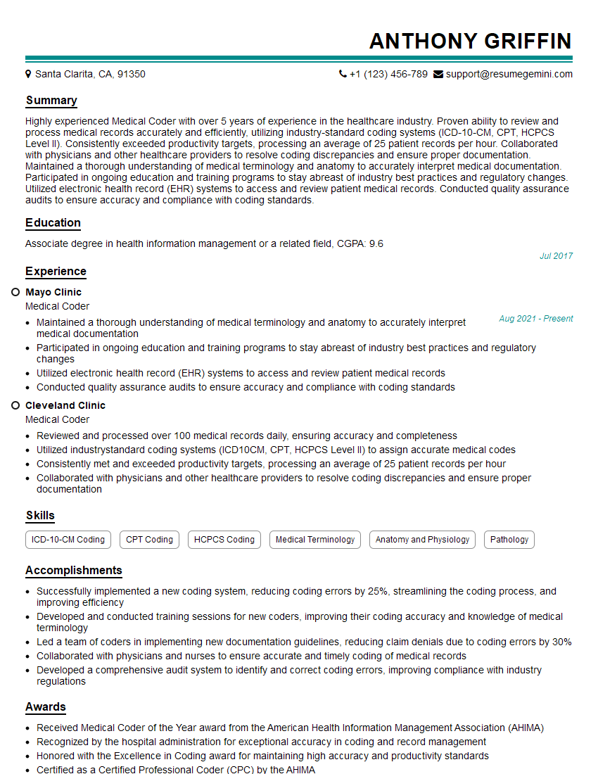
Mastering medical terminology and anatomy is vital for career advancement in healthcare. A strong understanding of these subjects demonstrates professionalism, attention to detail, and the ability to communicate effectively within a medical environment. To significantly improve your job prospects, create an ATS-friendly resume that highlights your skills and experience. ResumeGemini is a trusted resource that can help you build a professional and impactful resume. Examples of resumes tailored to showcasing expertise in medical terminology and anatomy are available, providing you with a valuable template for your own resume creation.
Explore more articles
Users Rating of Our Blogs
Share Your Experience
We value your feedback! Please rate our content and share your thoughts (optional).
What Readers Say About Our Blog
good
