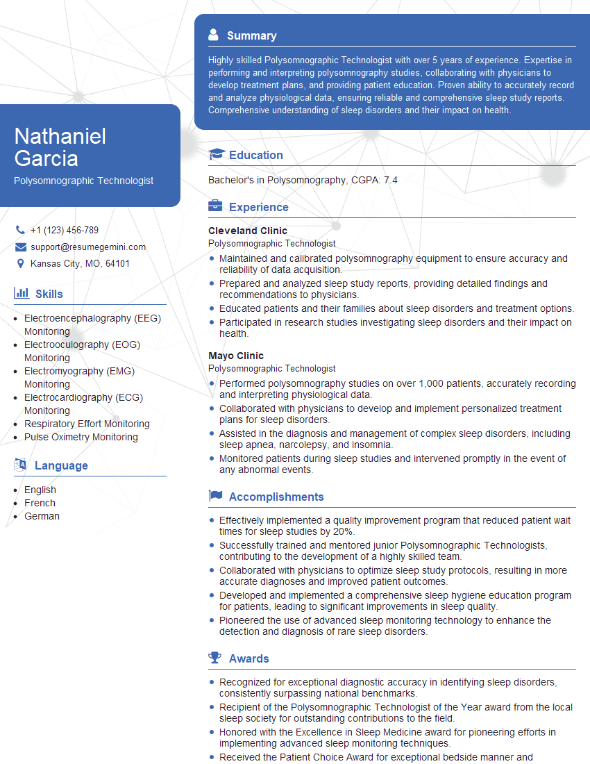
Are you ready to stand out in your next interview? Understanding and preparing for Physiological Monitoring interview questions is a game-changer. In this blog, we’ve compiled key questions and expert advice to help you showcase your skills with confidence and precision. Let’s get started on your journey to acing the interview.
Questions Asked in Physiological Monitoring Interview
Q 1. Explain the principles of ECG interpretation.
Electrocardiography (ECG) interpretation relies on understanding the electrical activity of the heart as reflected in the waveforms. Each waveform represents the depolarization (contraction) and repolarization (relaxation) of different parts of the heart. We analyze the ECG tracing for several key features:
- P wave: Represents atrial depolarization. A normal P wave is upright and smooth.
- QRS complex: Represents ventricular depolarization. This is the strongest deflection because ventricular muscle mass is much larger than atrial muscle mass.
- T wave: Represents ventricular repolarization. It’s typically upright but can be inverted in certain conditions.
- PR interval: Time from the start of atrial depolarization to the start of ventricular depolarization. It reflects the conduction time through the AV node.
- QRS duration: Duration of ventricular depolarization. Prolonged QRS duration can suggest a conduction delay.
- QT interval: Represents total ventricular depolarization and repolarization. This interval is crucial in assessing the risk of certain arrhythmias.
By analyzing the amplitude, duration, and morphology of these waveforms and intervals, we can diagnose a wide range of cardiac conditions, including arrhythmias, myocardial infarctions (heart attacks), electrolyte imbalances, and conduction abnormalities. For instance, a prolonged QT interval might indicate a risk of Torsades de Pointes, a potentially life-threatening arrhythmia. A widened QRS complex could signal a bundle branch block. Careful assessment of all components is essential for accurate interpretation.
Q 2. Describe the different types of arterial blood gas (ABG) analysis and their clinical significance.
Arterial blood gas (ABG) analysis measures the partial pressures of oxygen (PaO2) and carbon dioxide (PaCO2), pH, and bicarbonate (HCO3-) in arterial blood. This provides vital information about the respiratory and metabolic status of a patient. There are several types of ABG analysis, primarily differing in the methods used to collect and analyze the sample.
- Blood gas analyzer: This is the most common method, using electrochemical sensors to directly measure the components. Results are typically available within minutes.
- Point-of-care testing (POCT): Smaller, portable devices designed for rapid bedside testing, particularly useful in emergency situations. While less precise than laboratory analysis, they allow for faster decision making.
- Laboratory analysis: This involves sending the sample to a central lab for more detailed analysis which may include additional electrolytes and metabolic parameters.
The clinical significance of ABG analysis lies in its ability to diagnose and manage a wide range of conditions, including:
- Respiratory acidosis/alkalosis: Related to CO2 levels and ventilation.
- Metabolic acidosis/alkalosis: Related to bicarbonate levels and acid-base balance.
- Hypoxemia/Hypercapnia: Low oxygen and high carbon dioxide levels in the blood respectively, indicating respiratory impairment.
For example, a patient with severe pneumonia might present with hypoxemia (low PaO2) and hypercapnia (high PaCO2), indicating respiratory failure. ABG analysis is crucial for guiding oxygen therapy and ventilation strategies.
Q 3. What are the common causes of artifact in physiological monitoring?
Artifacts in physiological monitoring are unwanted signals that interfere with the accurate interpretation of the actual physiological data. These can originate from a variety of sources. Common causes include:
- Patient movement: This is the most frequent cause, especially with ECG and pulse oximetry. Muscle movement, shivering, or patient restlessness can distort waveforms.
- Electromagnetic interference (EMI): From nearby electronic devices like cell phones, electrical equipment, or even fluorescent lights. This can lead to noise and artifacts in ECG, EEG, and other signals.
- Loose or improperly placed sensors: Poor contact between the sensor and the patient’s skin can create artifacts, such as motion artifacts in ECG and pulse oximetry.
- Improper grounding: Inadequate grounding of equipment can create electrical noise and interfere with signals.
- Internal electrical activity: In ECG, high-frequency electrical activity from other sources in the patient’s body, such as muscle movements, can be mistaken for cardiac activity.
Recognizing and mitigating these artifacts is essential for obtaining reliable physiological data. For example, using proper grounding techniques, shielding equipment, and ensuring secure sensor placement can significantly reduce artifact contamination.
Q 4. How do you troubleshoot a malfunctioning pulse oximeter?
Troubleshooting a malfunctioning pulse oximeter involves a systematic approach. Here’s a step-by-step guide:
- Check sensor placement: Ensure the sensor is properly positioned on a fingertip or earlobe with good skin contact. Poor placement is a common source of error.
- Inspect the sensor and probe: Look for any damage to the sensor, such as cracks or dirt. Clean it with a suitable solution if necessary.
- Check the connection: Verify that the sensor is securely connected to the pulse oximeter and the pulse oximeter itself is properly connected to the power source.
- Examine the display: Check for error messages on the display. These may provide clues about the malfunction.
- Assess the patient’s condition: If the patient has poor peripheral perfusion (e.g., due to cold extremities or low blood pressure), this might interfere with accurate readings.
- Try a different sensor or probe: If possible, switch to a different sensor to see if the problem is with the sensor or the pulse oximeter itself.
- Check the ambient light: Excessive ambient light can interfere with readings; reduce the light if possible.
- Calibration: Some pulse oximeters require periodic calibration. Consult the device’s instructions to perform a calibration.
- Seek technical assistance: If the problem persists, contact the manufacturer or biomedical engineering department for assistance.
A systematic approach will quickly identify the issue and correct it, or escalate appropriately for repair or replacement.
Q 5. Explain the difference between invasive and non-invasive blood pressure monitoring.
The primary difference between invasive and non-invasive blood pressure monitoring lies in how the blood pressure is measured and the degree of patient intervention involved.
- Non-invasive blood pressure monitoring (NIBP): This is the most common method, utilizing a sphygmomanometer (blood pressure cuff) and stethoscope (auscultatory method) or an automated device (oscillometric method). The cuff is inflated to occlude blood flow, and then gradually deflated, with blood pressure determined by Korotkoff sounds (auscultatory) or pressure oscillations (oscillometric). It’s simple, relatively painless, and safe, but readings can be affected by factors like patient movement or improper cuff placement.
- Invasive blood pressure monitoring (IBP): This involves inserting a catheter into an artery (usually radial or femoral), connecting it to a transducer that measures blood pressure continuously. IBP provides real-time, beat-to-beat blood pressure readings, offering greater accuracy and continuous monitoring. However, it’s an invasive procedure carrying risks of infection, hemorrhage, thrombosis (blood clot formation), and nerve damage. It is reserved for critically ill patients needing continuous hemodynamic monitoring.
In summary, NIBP is preferred for routine blood pressure measurements, while IBP is reserved for situations requiring close monitoring of hemodynamic stability, such as in intensive care units (ICUs).
Q 6. What are the normal ranges for vital signs (heart rate, blood pressure, respiratory rate, SpO2, temperature)?
Normal ranges for vital signs vary slightly depending on factors such as age, sex, and overall health. However, general guidelines are as follows:
- Heart Rate (HR): 60-100 beats per minute (bpm) for adults. Children and infants have higher normal ranges.
- Blood Pressure (BP): Generally, less than 120/80 mmHg (systolic/diastolic) is considered ideal for adults. Ranges vary with age and individual health.
- Respiratory Rate (RR): 12-20 breaths per minute (bpm) for adults. Children and infants have higher normal ranges.
- SpO2 (Oxygen Saturation): 95-100% is considered normal. Lower values indicate hypoxemia (low blood oxygen).
- Temperature: Normal oral temperature is typically around 98.6°F (37°C), though individual variation exists. Rectal temperature is slightly higher.
It is crucial to remember that these are general ranges and individual variations exist. Deviations from these ranges necessitate further evaluation to determine the cause and appropriate interventions.
Q 7. Describe the process of applying and monitoring an arterial line.
Applying and monitoring an arterial line is a complex procedure requiring sterile technique and careful attention to detail. It should only be performed by trained healthcare professionals.
- Site selection: The most common sites are the radial, femoral, and brachial arteries. The site is assessed for suitability (e.g., good pulse, no previous injury).
- Preparation: The site is thoroughly cleaned with antiseptic solution, and sterile drapes are applied. Local anesthesia is usually administered.
- Arterial puncture: A small incision is made in the skin, and a needle is inserted into the artery. Blood return is confirmed before inserting the catheter.
- Catheter insertion: The catheter is advanced into the artery under guidance from pulse waveform or imaging in some cases. The needle is withdrawn, and the catheter is secured in place with sutures and a dressing.
- Transducer connection: The catheter is connected to a pressure transducer, which converts the arterial pressure waveform into an electrical signal that is displayed on a monitor.
- Waveform verification: The displayed waveform is checked for accuracy and appropriate characteristics (e.g., sharp upstroke, dicrotic notch).
- Continuous monitoring: Arterial blood pressure is continuously monitored. The site is closely observed for bleeding, infection, and hematoma formation.
- Regular maintenance: The arterial line requires regular flushing with heparinized saline to maintain patency and prevent clotting.
- Removal: When no longer needed, the arterial line is removed under sterile conditions. Pressure is applied to the puncture site to prevent bleeding.
Complications can include bleeding, infection, thrombosis, nerve injury, and air embolism. Meticulous technique and continuous monitoring are essential for minimizing these risks.
Q 8. How do you interpret a capnography waveform?
Capnography is a non-invasive method of monitoring ventilation by measuring the concentration of carbon dioxide (CO2) in the respiratory gases. The waveform, displayed on a capnograph, provides valuable information about the patient’s respiratory status.
A normal capnography waveform shows a characteristic rectangular shape. The waveform rises rapidly during exhalation as CO2 is expelled from the lungs, plateaus briefly, and then sharply declines as fresh gas enters the airways. Key aspects to interpret include:
- Baseline: Should be near zero, indicating absence of CO2 in the inspired air.
- Upstroke (Phase II): Represents the concentration of CO2 in the exhaled breath. A slow rise might suggest airway obstruction.
- Plateau (Phase III): The flat portion, indicating consistent CO2 concentration. A decreased plateau indicates hypoventilation.
- Downstroke (Phase IV): The rapid fall as the breath is completed. An abrupt downward slope is normal, a gradual slope might signify airway obstruction.
- End-tidal CO2 (EtCO2): The highest CO2 concentration at the end of exhalation. Normal values range from 35-45 mmHg. A significant increase or decrease can indicate serious respiratory problems.
Examples: A low EtCO2 might indicate hypoventilation, pulmonary embolism, or cardiac arrest. An elevated EtCO2 might suggest hyperventilation, rebreathing, or airway obstruction. A flatline indicates no exhaled CO2, signifying apnea or disconnection from the capnograph.
Q 9. Explain the significance of ST segment changes on an ECG.
The ST segment on an electrocardiogram (ECG) represents the early repolarization phase of the ventricles. Changes in the ST segment are extremely significant, often indicating myocardial ischemia (reduced blood flow to the heart muscle) or injury.
ST segment elevation (STE): Indicates acute myocardial infarction (heart attack), a life-threatening event where a portion of the heart muscle is dying due to lack of oxygen. The elevation is caused by the injured heart muscle depolarizing abnormally. The location of the STE on the ECG helps to pinpoint the location of the infarct.
ST segment depression (STD): Suggests myocardial ischemia, where the heart muscle is not receiving enough oxygen. This can be a sign of angina (chest pain), a warning sign of a possible future heart attack. STD may also be a result of other factors like electrolyte imbalances.
ST segment changes are not always indicative of heart problems. They can also be influenced by factors such as electrolyte disturbances, medication effects, and bundle branch blocks. It’s crucial for a cardiologist to analyze these changes within the context of the patient’s clinical presentation and other diagnostic tests.
Q 10. What are the signs and symptoms of hypoxemia and hypercapnia?
Hypoxemia is low blood oxygen levels, while hypercapnia is high blood carbon dioxide levels. Both can severely impact the body’s functions.
Hypoxemia Signs and Symptoms:
- Early signs: Increased respiratory rate, shortness of breath, and mild confusion or anxiety.
- Late signs: Cyanosis (bluish discoloration of the skin and mucous membranes), altered mental status, cardiac arrhythmias, and decreased blood pressure. In severe cases, loss of consciousness and organ failure.
Hypercapnia Signs and Symptoms:
- Early signs: Increased respiratory rate (often initially), headache, drowsiness, confusion, and slight tremors.
- Late signs: Lethargy, disorientation, coma, and potentially fatal cardiac arrhythmias. The body tries to compensate for elevated CO2 by increasing ventilation.
The severity of symptoms depends on the extent and speed of change in oxygen and carbon dioxide levels. It is important to remember that some patients, particularly those with chronic respiratory diseases, may have compensated for chronically low oxygen levels and may not show the typical symptoms.
Q 11. Describe the different types of cardiac arrhythmias and their treatment.
Cardiac arrhythmias are irregularities in the heart’s rhythm. They can be caused by problems in the heart’s electrical conduction system. There are many types, categorized by their origin (atria or ventricles) and rate (bradycardia or tachycardia).
Types and Treatments:
- Atrial fibrillation (AFib): Irregular and rapid heartbeat originating in the atria. Treatment includes rate control medications (e.g., beta-blockers), rhythm control medications (e.g., amiodarone), or cardioversion (to restore a normal rhythm).
- Atrial flutter: Rapid and regular heartbeat originating in the atria. Treatment is similar to AFib.
- Ventricular tachycardia (V-tach): Rapid heartbeat originating in the ventricles. Can be life-threatening. Treatment includes cardioversion, antiarrhythmic medications (e.g., amiodarone, lidocaine), or implantable cardioverter-defibrillator (ICD).
- Ventricular fibrillation (V-fib): Chaotic and irregular heartbeat originating in the ventricles. A life-threatening emergency requiring immediate defibrillation.
- Bradycardia: Slow heart rate. Treatment depends on the cause and may involve medications (e.g., atropine) or a pacemaker.
Treatment is tailored to the specific arrhythmia, its severity, and the patient’s overall health. It can involve medications, implantable devices (pacemakers, ICDs), or surgical procedures such as catheter ablation.
Q 12. How do you manage a patient with a sudden drop in blood pressure?
A sudden drop in blood pressure (hypotension) is a serious medical emergency requiring immediate action. The approach involves a systematic assessment and prompt treatment.
Management Steps:
- Assess the patient: Check vital signs (heart rate, respiratory rate, oxygen saturation), level of consciousness, and skin perfusion. Look for signs of shock (pale, clammy skin, rapid weak pulse).
- Identify the cause: Determine the underlying cause, such as bleeding, dehydration, heart failure, or sepsis.
- Position the patient: Place the patient in a supine position (lying flat on their back) with their legs elevated to increase blood flow to the brain.
- Administer fluids: If dehydration is suspected, administer intravenous fluids to increase blood volume.
- Treat the underlying cause: Address the root cause of the hypotension. This might involve stopping bleeding, administering antibiotics for sepsis, or medication for heart failure.
- Monitor vital signs: Closely monitor the patient’s blood pressure, heart rate, and other vital signs.
- Administer medications: Depending on the cause, medications such as vasopressors (to raise blood pressure) may be necessary.
Example: If a patient experiencing a sudden drop in blood pressure is bleeding profusely, immediate attention to controlling the bleeding is vital, alongside fluid resuscitation and potentially blood transfusions.
Q 13. What are the safety precautions when using a defibrillator?
Defibrillators deliver a high-energy electrical shock to the heart to stop life-threatening arrhythmias like ventricular fibrillation (V-fib) and pulseless ventricular tachycardia (V-tach).
Safety Precautions:
- Ensure the defibrillator is properly charged: Confirm that the device is correctly charged and ready to deliver a shock.
- Check the pads: Inspect the pads to ensure they are intact and properly applied to the patient’s chest, avoiding placing them over implanted devices if possible.
- Make sure the area is dry: Ensure the patient’s chest is dry to prevent burns and ensure optimal electrical conductivity.
- Clear the patient: Before delivering a shock, verbally shout “Clear!” to ensure no one is touching the patient.
- Deliver the shock: Once everyone is clear, press the shock button to deliver the defibrillator shock.
- Perform CPR: After delivering the shock, immediately begin cardiopulmonary resuscitation (CPR) until the patient’s spontaneous circulation returns or advanced medical care arrives.
- Do not touch the patient during shock delivery: Touching the patient during the shock delivery will expose oneself to the electric current, causing harm.
- Follow the manufacturer’s instructions: Always adhere to the defibrillator’s operating instructions.
Proper training and adherence to these safety measures are crucial to ensure both patient and operator safety during defibrillation.
Q 14. Explain the principles of ventilatory support.
Ventilatory support refers to providing assistance or taking over the function of breathing for patients who are unable to breathe adequately on their own. This can range from simple oxygen supplementation to advanced mechanical ventilation.
Principles:
- Oxygen Delivery: Increasing the amount of oxygen available to the patient, often by using supplemental oxygen via a mask or nasal cannula.
- Tidal Volume (Vt): The amount of air delivered to the lungs with each breath. Optimal Vt varies by patient needs and condition. Too much volume can cause lung injury (volutrauma), too little can lead to poor gas exchange.
- Respiratory Rate (RR): The number of breaths per minute. The rate is adjusted to maintain adequate gas exchange and minimize work of breathing.
- Positive End-Expiratory Pressure (PEEP): Positive pressure in the lungs at the end of exhalation. PEEP helps to keep the alveoli open, improving oxygenation.
- Inspiratory Pressure Support: Assisting the patient’s own inspiratory effort by providing additional pressure during inhalation.
- Mode Selection: Different modes of ventilation exist, such as assist-control ventilation (where the ventilator delivers a breath every set time) or pressure support ventilation (where the ventilator supports the patient’s breaths as needed). Selection depends on the patient’s condition and needs.
- Monitoring: Continuous monitoring of vital signs and ventilator settings are crucial to ensure effective ventilation and minimize complications. This includes close monitoring of blood gas analysis (ABGs).
Ventilatory support requires specialized training and knowledge. Careful management is essential to prevent complications like barotrauma (lung injury from pressure), volutrauma, and ventilator-associated pneumonia.
Q 15. Describe the different modes of mechanical ventilation.
Mechanical ventilation supports breathing by delivering oxygen and removing carbon dioxide. Different modes adjust the ventilator’s contribution based on the patient’s respiratory effort. Common modes include:
- Volume-controlled ventilation (VCV): The ventilator delivers a preset tidal volume (the amount of air per breath) at a set respiratory rate. The pressure required to deliver this volume varies depending on lung compliance and airway resistance. This is often used for patients who are unable to initiate breaths on their own.
- Pressure-controlled ventilation (PCV): The ventilator delivers a preset pressure for a set inspiratory time. The tidal volume varies depending on lung compliance and airway resistance. This can be gentler on the lungs than VCV.
- Assist-control ventilation (ACV): The ventilator delivers a breath at a set rate and tidal volume but also delivers breaths triggered by the patient’s own respiratory effort. This allows for patient participation in breathing.
- Synchronized intermittent mandatory ventilation (SIMV): The ventilator delivers a set number of breaths per minute (mandatory breaths) synchronized with the patient’s own breathing. The patient can breathe spontaneously between the ventilator breaths.
- Pressure support ventilation (PSV): The ventilator provides pressure support to help the patient breathe spontaneously. This mode is often used during weaning from mechanical ventilation.
- Non-invasive ventilation (NIV): Includes techniques like CPAP (continuous positive airway pressure) and BiPAP (bilevel positive airway pressure), delivered via a mask and used to support breathing without an endotracheal tube. This is beneficial in cases of respiratory failure, COPD exacerbation or sleep apnea.
The choice of ventilation mode depends on the patient’s condition, respiratory drive, and lung mechanics. For instance, a patient with acute respiratory distress syndrome (ARDS) might initially require VCV to ensure adequate oxygenation, while a patient recovering from surgery may transition to PSV to promote spontaneous breathing.
Career Expert Tips:
- Ace those interviews! Prepare effectively by reviewing the Top 50 Most Common Interview Questions on ResumeGemini.
- Navigate your job search with confidence! Explore a wide range of Career Tips on ResumeGemini. Learn about common challenges and recommendations to overcome them.
- Craft the perfect resume! Master the Art of Resume Writing with ResumeGemini’s guide. Showcase your unique qualifications and achievements effectively.
- Don’t miss out on holiday savings! Build your dream resume with ResumeGemini’s ATS optimized templates.
Q 16. How do you interpret a central venous pressure (CVP) waveform?
The central venous pressure (CVP) waveform reflects right atrial pressure and indirectly reflects right ventricular preload and fluid status. A normal CVP waveform has distinct components:
- a wave: Represents atrial contraction. A prominent ‘a’ wave can suggest tricuspid stenosis or right atrial hypertension.
- c wave: Reflects ventricular contraction and tricuspid valve bulging into the atrium. An exaggerated ‘c’ wave may indicate tricuspid regurgitation.
- x descent: Caused by atrial relaxation and downward displacement of the tricuspid valve.
- v wave: Represents venous return filling the right atrium while the tricuspid valve is closed.
- y descent: Represents passive emptying of the right atrium as the tricuspid valve opens.
Interpreting the waveform requires considering the clinical context. For example, a low CVP might suggest hypovolemia (low blood volume), while an elevated CVP might indicate fluid overload, right ventricular failure, or cardiac tamponade. It is crucial to correlate the CVP waveform with other clinical parameters like blood pressure, heart rate, and urine output for accurate interpretation. Analyzing the waveform alone is not enough; a comprehensive assessment of the patient is required.
Q 17. What are the potential complications of invasive hemodynamic monitoring?
Invasive hemodynamic monitoring, while providing valuable data, carries potential complications:
- Infection: Catheter-related bloodstream infections are a significant risk, particularly with central venous catheters. Meticulous sterile technique during insertion and diligent care are vital to prevent this.
- Bleeding: Puncture sites can bleed, especially in patients on anticoagulants. Careful monitoring and pressure dressings are essential.
- Thrombosis: Blood clots can form at the catheter insertion site or within the catheter itself. Regular flushing and careful catheter manipulation can help reduce this risk.
- Air embolism: Air can enter the bloodstream during catheter insertion or manipulation. This is a life-threatening complication requiring immediate attention.
- Pneumothorax: Accidental lung puncture during central line insertion can cause a pneumothorax. Chest x-rays are usually performed post-insertion to check for this.
- Catheter malposition: The catheter can be inadvertently placed in the wrong vessel, leading to inaccurate readings or damage to blood vessels.
The benefits of invasive hemodynamic monitoring must always be weighed against the potential risks. Careful patient selection, meticulous technique, and diligent post-insertion monitoring are crucial to minimize complications.
Q 18. Explain the importance of accurate documentation in physiological monitoring.
Accurate documentation in physiological monitoring is paramount for several reasons:
- Legal protection: Detailed records serve as legal evidence of the care provided. This is crucial for protecting both the patient and the healthcare provider.
- Continuity of care: Accurate documentation ensures seamless transfer of information among healthcare professionals, leading to better patient outcomes.
- Clinical decision-making: Comprehensive data allows for better clinical decision-making. Trends in physiological parameters can help identify deterioration early on.
- Research and quality improvement: Data collected through accurate documentation contributes to research and helps identify areas for quality improvement.
- Patient safety: Clear documentation minimizes the risk of medication errors or treatment omissions.
Documentation should include date, time, type of monitoring, specific readings, any interventions made, and the patient’s response. In a real-world scenario, for example, documenting the precise time a patient’s oxygen saturation dropped, along with the interventions taken (e.g., increasing oxygen flow, repositioning), is crucial for tracking the patient’s progress and assessing the effectiveness of treatment.
Q 19. How do you communicate effectively with a multidisciplinary team during a critical event?
Effective communication during a critical event in a multidisciplinary team requires clear, concise, and structured communication. I utilize the SBAR (Situation, Background, Assessment, Recommendation) framework:
- Situation: Briefly state the problem – e.g., ‘The patient’s blood pressure has dropped to 70/40 mmHg.’
- Background: Provide relevant context – e.g., ‘The patient is post-operative, and has been experiencing increased bleeding.’
- Assessment: State your assessment of the situation – e.g., ‘I suspect hypovolemic shock.’
- Recommendation: State what action needs to be taken – e.g., ‘I recommend immediate fluid resuscitation and contacting the surgical team.’
I also believe in active listening, seeking clarification, and ensuring everyone understands the plan. Using clear, non-medical jargon when addressing family members is equally critical in such scenarios. For example, instead of saying ‘tachycardia’, I would say ‘rapid heart rate’. The key is to ensure everyone, regardless of their background, is informed and involved in the decision-making process.
Q 20. Describe your experience with different types of physiological monitoring equipment.
My experience encompasses a wide range of physiological monitoring equipment, including:
- Cardiac monitors: I am proficient with various models, capable of interpreting ECG rhythms, ST segment changes, and other cardiac parameters. I have experience with both bedside and central monitoring systems.
- Hemodynamic monitoring systems: I have extensive experience with arterial line, central venous pressure (CVP) catheters, pulmonary artery catheters (PACs) and their associated equipment. This includes understanding the waveforms, troubleshooting equipment malfunctions and interpreting the data.
- Pulse oximeters: I am well-versed in using and interpreting pulse oximetry data, recognizing the limitations and potential sources of error.
- Capnography: I am proficient in interpreting capnography waveforms and using this data to assess ventilation.
- Mechanical ventilators: I have extensive experience working with various types of mechanical ventilators, understanding different modes, settings, and alarms.
- Non-invasive blood pressure monitors: I can correctly use and troubleshoot automated and manual blood pressure cuffs.
I am adept at using and interpreting data from these devices to guide treatment decisions and recognize potential issues. Furthermore, I am familiar with quality assurance procedures for all equipment.
Q 21. How do you handle stressful situations in a fast-paced environment?
Fast-paced, high-pressure environments are inherent to critical care. My approach to handling stress involves a multi-pronged strategy:
- Prioritization: I focus on identifying and addressing the most critical issues first, using a systematic approach to manage multiple tasks concurrently.
- Teamwork: I rely on strong teamwork and collaboration to distribute workload and share the burden of responsibility.
- Situational awareness: I maintain situational awareness, constantly reassessing the patient’s condition and adapting my approach as needed.
- Self-care: Outside of work, I prioritize healthy lifestyle choices such as exercise, proper nutrition and sleep to manage stress levels effectively. I also utilize mindfulness techniques and stress management strategies.
- Debriefing: After critical events, I participate in debriefing sessions with colleagues to review events, identify areas for improvement, and process emotional responses. This promotes team cohesion and learning.
By employing these strategies, I can manage stressful situations effectively and maintain composure, enabling me to provide optimal patient care even in challenging circumstances.
Q 22. Explain the importance of patient safety in physiological monitoring.
Patient safety is paramount in physiological monitoring. It’s the cornerstone of ethical and effective healthcare. Accurate and timely monitoring directly impacts clinical decision-making, preventing adverse events and improving patient outcomes. Imagine a scenario where a patient’s oxygen saturation drops dangerously low – without continuous monitoring, this critical change might go unnoticed, leading to serious complications or even death. Therefore, our vigilance and precise readings are crucial to ensuring timely interventions.
This importance manifests in several ways: early detection of life-threatening conditions (e.g., cardiac arrhythmias, respiratory distress), accurate assessment of response to treatment, and prevention of medication errors by providing objective data for dosage adjustments. Our role extends beyond just reading numbers; it’s about understanding the patient’s overall clinical picture and alerting the healthcare team to significant changes promptly.
Q 23. Describe your knowledge of relevant regulatory guidelines and standards.
My knowledge of regulatory guidelines and standards is extensive. I am thoroughly familiar with AAMI (Association for the Advancement of Medical Instrumentation) standards, which define the performance requirements for medical equipment, including physiological monitors. I also adhere to FDA (Food and Drug Administration) regulations concerning device safety and efficacy. Additionally, I understand the importance of following hospital-specific protocols and policies concerning equipment calibration, maintenance, and alarm management. These regulations and standards are essential to ensure patient safety and the reliability of monitoring data. Failure to comply can have serious consequences, including legal liabilities.
For instance, AAMI standards guide us in choosing equipment appropriate for specific patient populations, maintaining its accuracy, and interpreting the resulting data correctly. Understanding FDA requirements ensures that we use only approved equipment and that these devices are used according to their intended purposes and safety guidelines.
Q 24. How do you maintain your competency in physiological monitoring?
Maintaining competency in physiological monitoring requires ongoing dedication. I achieve this through a multi-pronged approach:
- Continuing Education: I actively participate in workshops, conferences, and online courses focused on the latest advancements in monitoring technology and clinical best practices.
- Professional Organizations: Membership in professional organizations, such as AAMI, keeps me abreast of industry changes and provides access to educational resources and networking opportunities.
- Journal Articles and Research: I regularly review peer-reviewed journals and research papers to stay updated on the latest findings and innovations in physiological monitoring.
- Case Studies and Internal Training: We conduct regular internal training sessions where we analyze complex cases, discuss best practices, and refine our skills in troubleshooting and decision-making.
- Mentorship: I actively seek mentorship opportunities and share my experience with colleagues, fostering a culture of continuous learning.
This continuous learning ensures that I stay proficient in interpreting complex waveforms, managing alarms effectively, and applying my knowledge to optimize patient care in diverse clinical settings.
Q 25. What are your strengths and weaknesses as a physiological monitoring professional?
My strengths lie in my meticulous attention to detail, critical thinking skills, and ability to quickly assess and prioritize patient needs in high-pressure situations. I’m highly proficient in troubleshooting equipment and adept at communicating effectively with healthcare teams. I thrive in dynamic environments and am comfortable working independently as well as part of a larger team.
One area where I’m actively working on improvement is expanding my knowledge of the newer, more advanced types of monitoring technology, specifically those involving artificial intelligence in data analysis. Although I can use them, a deeper understanding of the algorithms and potential limitations will enhance my ability to interpret the data and assist clinicians more effectively. I actively seek out training and resources to address this.
Q 26. Describe your experience with troubleshooting and resolving equipment malfunctions.
Troubleshooting equipment malfunctions is a routine part of my work. My approach is systematic and follows a structured process:
- Identify the problem: I begin by carefully observing the monitor, noting specific error messages or unusual waveforms.
- Check the obvious: I verify that the sensors are properly connected, the power is on, and there are no loose cables.
- Consult the manual: I refer to the equipment’s user manual for troubleshooting guides and suggested solutions.
- Systematic testing: If the problem persists, I perform systematic checks – testing each component of the monitoring system (e.g., cables, sensors, and the monitor itself) to pinpoint the source of the malfunction.
- Escalation: If the issue remains unresolved, I escalate the problem to the biomedical engineering department for repair or replacement.
- Documentation: I meticulously document all troubleshooting steps and the final resolution, which aids in future problem-solving and maintains accurate records.
For example, recently, a patient’s ECG monitor showed a flatline. Through systematic checks, I identified a disconnected lead wire. Replacing the wire immediately resolved the issue, avoiding a potential delay in detecting a serious cardiac event. Effective troubleshooting is not only about technical skills; it’s also about quick thinking and the ability to prioritize patient safety.
Q 27. How do you prioritize tasks during a high-volume workload?
Prioritizing tasks during a high-volume workload requires a structured approach. I use a combination of techniques:
- ABC prioritization: I categorize tasks into A (urgent and critical), B (important but not urgent), and C (less important) categories. This ensures that life-threatening situations are addressed immediately.
- Time management: I use time management techniques such as time blocking and the Pomodoro Technique to focus on individual tasks and avoid feeling overwhelmed.
- Delegation: Where appropriate, I delegate tasks to other qualified team members to ensure efficient use of resources.
- Communication: I maintain clear communication with colleagues and healthcare providers to ensure that everyone is aware of the priorities and the current situation. This teamwork approach ensures that critical issues get the attention they need.
In a busy ICU, for example, I might prioritize a patient experiencing respiratory distress (A) over routine monitoring checks (B) while still ensuring all patients receive the necessary care. This constant assessment and adaptation ensure that patient needs are met effectively, even under pressure.
Q 28. Explain your understanding of the legal and ethical implications of physiological monitoring.
Physiological monitoring involves significant legal and ethical implications. Accuracy and reliability of data are crucial for informed clinical decision-making. Errors in monitoring or misinterpretation of data can have serious consequences, including legal liability for negligence. Ethical considerations encompass patient confidentiality, data security, and the responsible use of technology.
- Confidentiality: Patient data must be protected according to HIPAA and other relevant regulations. Access should be strictly limited to authorized personnel.
- Data Integrity: We are responsible for ensuring the accuracy and reliability of the monitoring data. Calibration, maintenance, and proper sensor application are critical aspects.
- Informed Consent: Patients should be informed about the purpose and procedures of physiological monitoring. Their consent must be obtained appropriately.
- Professional Responsibility: We have a professional obligation to use our expertise responsibly, adhering to ethical guidelines and reporting any concerns about data integrity or patient safety.
For instance, a breach of patient confidentiality, like discussing a patient’s vital signs with unauthorized individuals, could lead to disciplinary action. Similarly, failure to properly maintain equipment resulting in inaccurate readings could have legal repercussions.
Key Topics to Learn for Physiological Monitoring Interview
- Electrocardiography (ECG): Understanding ECG waveforms, arrhythmia recognition, and lead placement techniques. Practical application: Interpreting ECG strips to identify cardiac abnormalities and inform clinical decisions.
- Hemodynamic Monitoring: Mastering invasive and non-invasive blood pressure measurement, central venous pressure (CVP), pulmonary artery catheter (PAC) interpretation. Practical application: Titrating medications based on hemodynamic parameters to optimize patient care.
- Respiratory Monitoring: Understanding oxygen saturation (SpO2), capnography (EtCO2), and mechanical ventilation parameters. Practical application: Identifying respiratory distress and adjusting ventilator settings to improve gas exchange.
- Neurological Monitoring: Interpreting intracranial pressure (ICP) waveforms, electroencephalograms (EEGs), and other neurological data. Practical application: Assessing neurological status and adjusting treatment plans based on monitored data.
- Equipment Operation and Troubleshooting: Gaining hands-on experience with various monitoring devices and understanding common malfunctions and their solutions. Practical application: Quickly identifying and resolving equipment issues to ensure patient safety.
- Patient Safety and Communication: Understanding protocols for alarm management, recognizing critical situations, and effectively communicating with healthcare teams. Practical application: Prioritizing patient safety and collaborating effectively with colleagues.
- Legal and Ethical Considerations: Familiarizing yourself with relevant regulations and ethical guidelines related to patient privacy and data security. Practical application: Maintaining ethical practices and ensuring compliance with all regulations.
Next Steps
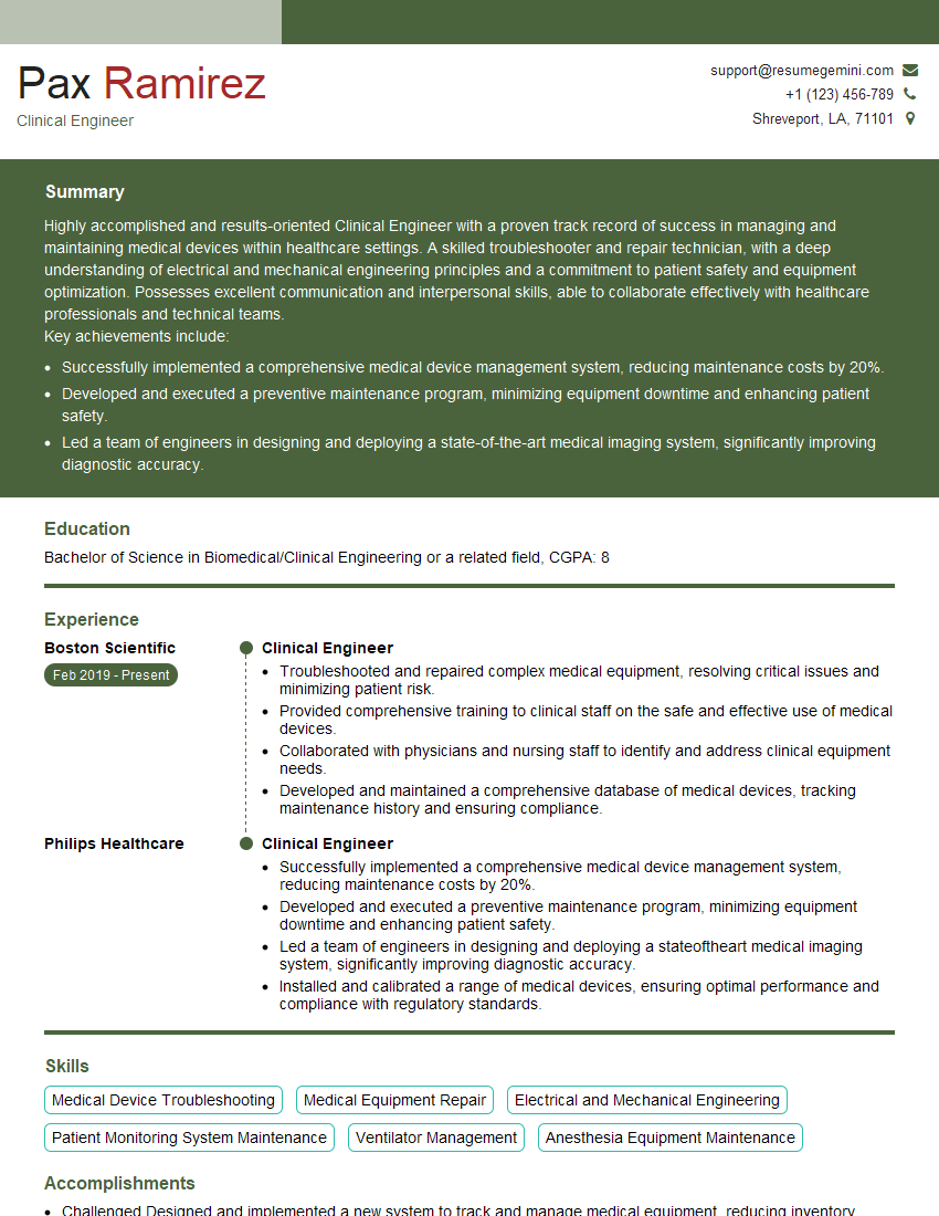
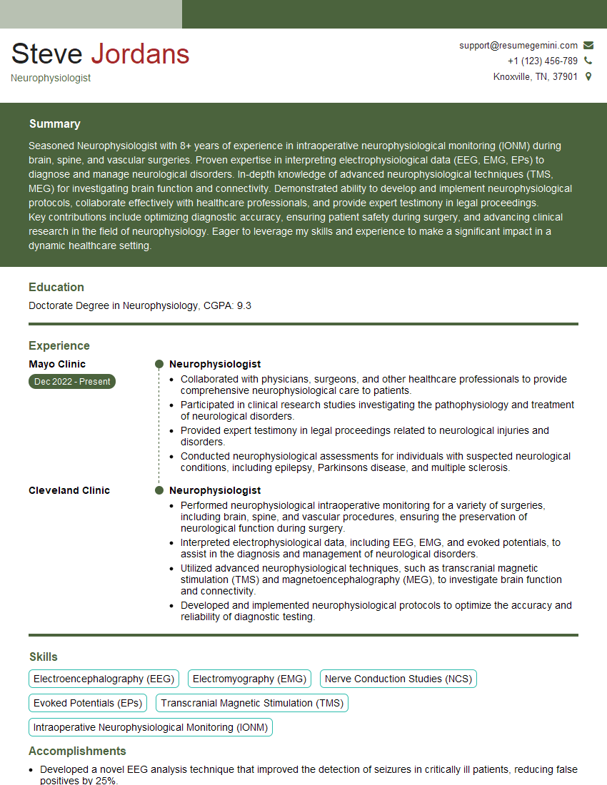
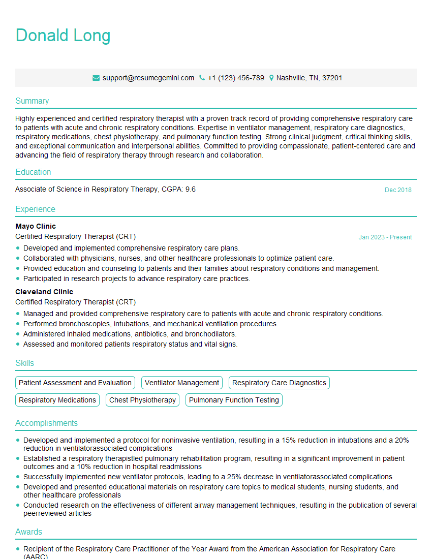
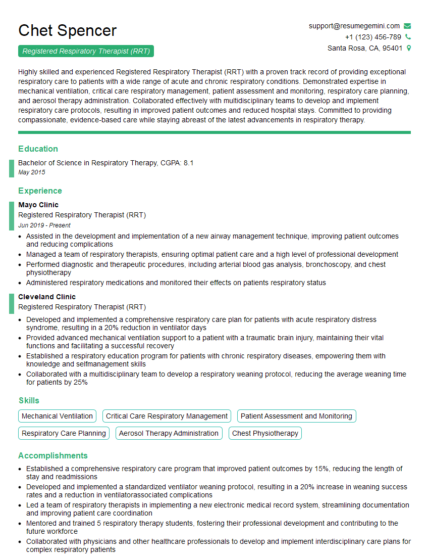
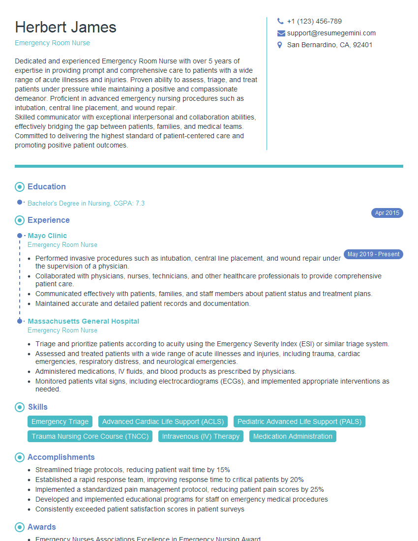
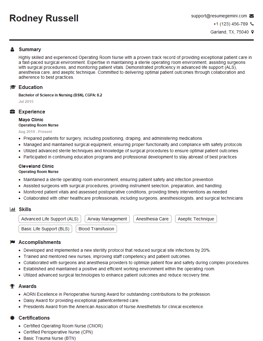
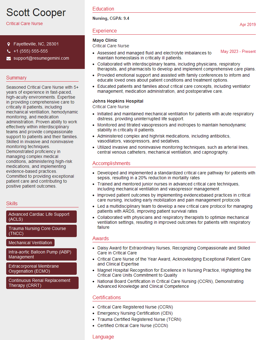
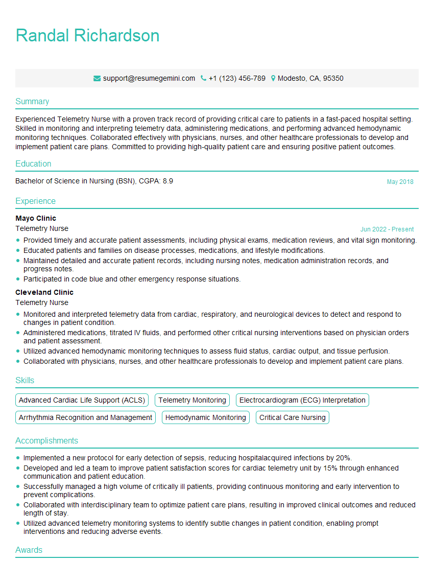
Mastering Physiological Monitoring opens doors to exciting career opportunities in critical care, operating rooms, and various specialized medical settings. It demonstrates a high level of technical expertise and commitment to patient well-being, making you a highly valuable asset to any healthcare team. To maximize your job prospects, crafting an ATS-friendly resume is crucial. ResumeGemini is a trusted resource that can help you build a professional and impactful resume that highlights your skills and experience. Examples of resumes tailored to Physiological Monitoring are available through ResumeGemini to guide you in showcasing your qualifications effectively.
Explore more articles
Users Rating of Our Blogs
Share Your Experience
We value your feedback! Please rate our content and share your thoughts (optional).
What Readers Say About Our Blog
Hello,
We found issues with your domain’s email setup that may be sending your messages to spam or blocking them completely. InboxShield Mini shows you how to fix it in minutes — no tech skills required.
Scan your domain now for details: https://inboxshield-mini.com/
— Adam @ InboxShield Mini
Reply STOP to unsubscribe
Hi, are you owner of interviewgemini.com? What if I told you I could help you find extra time in your schedule, reconnect with leads you didn’t even realize you missed, and bring in more “I want to work with you” conversations, without increasing your ad spend or hiring a full-time employee?
All with a flexible, budget-friendly service that could easily pay for itself. Sounds good?
Would it be nice to jump on a quick 10-minute call so I can show you exactly how we make this work?
Best,
Hapei
Marketing Director
Hey, I know you’re the owner of interviewgemini.com. I’ll be quick.
Fundraising for your business is tough and time-consuming. We make it easier by guaranteeing two private investor meetings each month, for six months. No demos, no pitch events – just direct introductions to active investors matched to your startup.
If youR17;re raising, this could help you build real momentum. Want me to send more info?
Hi, I represent an SEO company that specialises in getting you AI citations and higher rankings on Google. I’d like to offer you a 100% free SEO audit for your website. Would you be interested?
Hi, I represent an SEO company that specialises in getting you AI citations and higher rankings on Google. I’d like to offer you a 100% free SEO audit for your website. Would you be interested?
good
