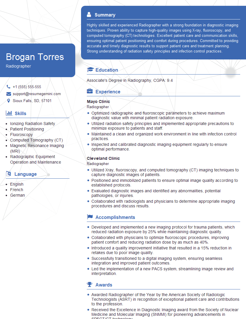
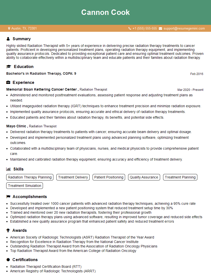
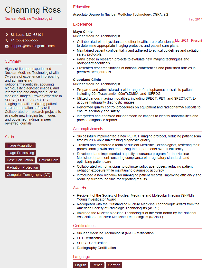
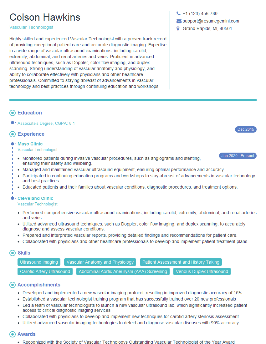
Cracking a skill-specific interview, like one for RT Certification, requires understanding the nuances of the role. In this blog, we present the questions you’re most likely to encounter, along with insights into how to answer them effectively. Let’s ensure you’re ready to make a strong impression.
Questions Asked in RT Certification Interview
Q 1. Explain the ALARA principle and its application in radiology.
The ALARA principle, short for “As Low As Reasonably Achievable,” is a fundamental guideline in radiation protection. It emphasizes minimizing radiation exposure to patients and healthcare workers while still achieving the diagnostic goals. It’s not about eliminating radiation entirely – that’s often impossible – but about reducing it to the lowest level that’s practical and justifiable.
In radiology, ALARA is applied through various methods. For example, optimizing exposure parameters during X-ray imaging (reducing the kVp and mAs while maintaining image quality), using proper shielding (lead aprons, thyroid collars), employing appropriate collimation techniques to restrict the radiation beam to the area of interest, and using image intensifiers with low radiation output during fluoroscopy.
Imagine taking a picture with your phone. You wouldn’t want to use the brightest flash possible all the time, would you? ALARA is similar; we want the clearest image possible with the least amount of radiation.
Q 2. Describe the different types of ionizing radiation used in medical imaging.
Medical imaging utilizes several types of ionizing radiation. The most common are:
- X-rays: Electromagnetic radiation used in conventional radiography, fluoroscopy, and computed tomography (CT). X-rays have high energy and can penetrate tissues, creating an image based on differential absorption.
- Gamma rays: High-energy electromagnetic radiation emitted by radioactive isotopes used in nuclear medicine procedures like SPECT and PET scans. Gamma rays are used to detect and image the distribution of radioactive tracers within the body.
- Electron beams: Used in radiation therapy, although not strictly medical imaging. They deliver a high dose of radiation to precisely target tumors.
The choice of radiation depends on the imaging modality and the specific diagnostic information needed. For example, X-rays are ideal for visualizing bone fractures, while gamma rays are better suited for evaluating metabolic activity within organs.
Q 3. What are the safety precautions for patients and healthcare workers during fluoroscopy procedures?
Fluoroscopy, a real-time X-ray imaging technique, requires stringent safety measures for both patients and healthcare workers. For patients, this includes:
- Minimizing exposure time: The fluoroscopy beam should only be activated when absolutely necessary. Pulse fluoroscopy, which intermittently emits radiation, helps reduce exposure.
- Using proper shielding: Lead aprons and thyroid collars should be used to protect sensitive organs from scatter radiation.
- Optimizing image parameters: Maintaining the lowest radiation output consistent with a diagnostically useful image.
For healthcare workers, safety measures include:
- Distance: Maintaining a safe distance from the radiation source during fluoroscopy procedures.
- Shielding: Using lead aprons, gloves, and glasses whenever possible.
- Time minimization: Limiting exposure time by assigning tasks efficiently and utilizing lead shielding.
- Rotation of personnel: Ensuring a consistent rotation of staff involved in fluoroscopy procedures to limit individual exposure.
Remember, even seemingly small exposures can accumulate over time, so strict adherence to safety protocols is crucial.
Q 4. How do you ensure proper patient positioning for various imaging modalities?
Proper patient positioning is critical for producing high-quality images across all imaging modalities. It ensures the anatomical structures of interest are optimally aligned with the radiation beam or magnetic field. The process involves a combination of anatomical knowledge, precise manipulation, and the use of positioning aids (such as wedges and sponges).
For example, in chest X-rays, proper positioning means ensuring the patient is erect, their shoulders rolled forward, and their arms are positioned away from the lungs to prevent obscuring lung fields. In MRI, accurate patient positioning within the scanner coil is critical for obtaining sharp and artifact-free images. Each modality has specific protocols for positioning, dependent upon the area of the body being imaged and the required anatomical view.
My experience involves ensuring patients are comfortable and understanding the rationale for positioning. Often, a simple explanation of the procedure and its importance can alleviate patient anxiety and assist in achieving the correct positioning.
Q 5. What are the differences between CT, MRI, and X-ray imaging?
CT, MRI, and X-ray imaging are all crucial medical imaging techniques, but they differ significantly in their principles and applications:
- X-ray imaging: Uses ionizing radiation (X-rays) to produce two-dimensional images of the body. It’s excellent for visualizing bone, but soft tissues are less well-defined.
- CT (Computed Tomography): Employs X-rays to generate cross-sectional images of the body. It provides much better visualization of soft tissue than conventional X-rays and allows for 3D reconstructions. It’s widely used for trauma assessment, cancer detection, and guidance during procedures.
- MRI (Magnetic Resonance Imaging): Uses strong magnetic fields and radio waves to create detailed images of the body’s internal structures. It excels in visualizing soft tissues, such as the brain, spinal cord, and ligaments. It doesn’t use ionizing radiation, making it safer for repeated scans.
In short: X-rays are quick and readily available for basic imaging, CT offers high-resolution cross-sectional views, and MRI provides excellent soft-tissue contrast without ionizing radiation.
Q 6. Explain the process of image acquisition and processing in digital radiography.
Digital radiography (DR) involves image acquisition and processing steps significantly different from traditional film-based radiography. The process begins with X-ray photons interacting with the patient’s body. The resulting image is captured by a digital detector (flat-panel detector or CCD).
Image Acquisition: The X-ray detector measures the intensity of the transmitted X-rays, converting the analog signal into a digital signal, which is represented as a matrix of pixel values.
Image Processing: The digital data undergoes several processing steps to enhance image quality. These steps include:
- Signal amplification: Increasing the signal strength to improve image visibility.
- Noise reduction: Removing unwanted artifacts to improve image clarity.
- Image scaling and contrast adjustment: Optimizing image brightness and contrast for better visualization.
- Image compression: Reducing file size for efficient storage and transmission.
The final processed image is then displayed on a monitor and can be stored, transmitted, and archived using a PACS system.
Q 7. Describe your experience with PACS (Picture Archiving and Communication Systems).
I have extensive experience working with PACS (Picture Archiving and Communication Systems). PACS systems are crucial for efficient management of medical images, allowing for centralized storage, retrieval, and distribution of images across different modalities. My experience includes:
- Image archiving and retrieval: Storing and retrieving medical images efficiently and securely, using appropriate naming conventions and archiving protocols.
- DICOM standards: Working with DICOM (Digital Imaging and Communications in Medicine) standards, ensuring seamless integration and communication between imaging modalities and the PACS.
- Image viewing and manipulation: Utilizing PACS software to view, annotate, and manipulate medical images, improving diagnostics and collaboration.
- Troubleshooting: Identifying and resolving PACS-related technical issues to maintain system uptime and workflow.
- System administration: Contributing to the maintenance and upgrade of PACS infrastructure to ensure its optimal performance.
I’m proficient in using various PACS systems and understand the importance of maintaining data integrity and security within these systems. This includes secure access control and regular data backups to protect patient data.
Q 8. How do you handle a malfunctioning imaging machine?
Handling a malfunctioning imaging machine requires a calm and systematic approach, prioritizing patient safety and equipment integrity. First, I would ensure the patient is safe and remove them from the immediate vicinity if necessary. Then, I would follow established protocols for equipment malfunction, which typically involves turning off the machine at the main power switch.
Next, I would report the malfunction immediately to the designated personnel, such as a biomedical engineer or supervisor. This is crucial for timely repair and prevents further incidents. The specific steps might vary slightly depending on the type of machine (e.g., X-ray, CT, MRI) but generally involve documenting the malfunction, including the time, nature of the problem, and any relevant details. Finally, I would ensure the area is secure and avoid using the malfunctioning machine until it has been inspected and repaired by qualified personnel. Think of it like this: you wouldn’t drive a car with a flat tire; you’d report it and have it fixed. The same applies to medical imaging equipment.
For example, if an X-ray machine fails to produce an image, I’d check the power supply and exposure settings, then contact the biomedical engineer and document the error in the machine’s logbook. If a CT scanner stops mid-scan, I’d immediately stop the scan, assess the patient’s condition, and notify the radiologist and biomedical engineer immediately.
Q 9. What are the legal and ethical responsibilities of a radiologic technologist?
A radiologic technologist’s legal and ethical responsibilities are paramount. Legally, we must adhere to all relevant state and federal regulations regarding radiation safety, patient privacy (HIPAA), and medical record keeping. This includes accurate documentation of procedures, proper patient identification, and strict adherence to radiation safety protocols. Failure to do so can result in legal repercussions, including lawsuits and loss of licensure. Ethically, we have a responsibility to provide the highest quality patient care, treating every patient with respect, dignity, and compassion. This involves maintaining patient confidentiality, accurately interpreting and communicating imaging findings, and participating in continuing education to stay current on best practices.
For instance, we are ethically obligated to advocate for our patients, ensuring they understand the procedure and have the opportunity to ask questions before proceeding. We must also remain objective, avoiding any bias in our assessment of imaging results and reporting them to the physician for accurate diagnosis.
Q 10. Explain the importance of radiation safety and protection.
Radiation safety and protection are absolutely critical in radiology. Ionizing radiation, while useful for medical imaging, carries the risk of causing harm to living tissues, potentially leading to long-term health problems like cancer. Therefore, minimizing radiation exposure is a top priority. This involves using the ALARA principle – As Low As Reasonably Achievable – in all aspects of imaging procedures.
This includes optimizing imaging parameters (e.g., minimizing exposure time, using appropriate kVp and mAs settings), utilizing proper shielding techniques (e.g., lead aprons, thyroid shields, collimators), and implementing appropriate safety protocols. Patient education is also crucial. Patients should understand the benefits and risks of radiation exposure before undergoing any procedure. We use various radiation monitoring devices to ensure compliance with safety regulations, such as personal dosimeters and radiation area monitors. Regular maintenance checks on imaging equipment are vital to prevent malfunctions that could result in increased radiation exposure. Think of it like wearing sunscreen to protect yourself from harmful UV rays – we use multiple layers of protection to shield patients and ourselves from radiation.
Q 11. How do you maintain patient confidentiality?
Maintaining patient confidentiality is a cornerstone of ethical and legal practice in healthcare. This is primarily governed by the Health Insurance Portability and Accountability Act (HIPAA) in the United States. In practice, this means never discussing patient information with unauthorized individuals. This includes family members, friends, or even other healthcare professionals not directly involved in the patient’s care. I strictly adhere to HIPAA regulations, ensuring that all patient information is kept secure, whether in electronic or physical form. Access to patient files is restricted to authorized personnel only and I use secure passwords and encryption for electronic medical records. I also ensure all conversations about patients are held in private areas. Additionally, I am very mindful of leaving patient information unsecured, like on a computer screen that is visible to passersby.
Imagine a bank; they take strict measures to safeguard your financial information. Similarly, we must treat patient health information with the utmost care and discretion.
Q 12. Describe your experience with various contrast media and their administration.
I have extensive experience with various contrast media, including ionic and non-ionic iodinated contrast agents, as well as gadolinium-based contrast agents used in MRI. I am proficient in the safe administration of these agents, understanding their indications, contraindications, and potential adverse reactions. Before administering any contrast, I meticulously review the patient’s medical history to identify potential allergies or contraindications. This includes assessing renal function (especially crucial for iodinated contrast). I carefully monitor patients during and after contrast administration for any signs of allergic reactions (e.g., hives, swelling, shortness of breath) and am trained to manage these reactions should they occur. For example, I am familiar with the protocols for administering medications like antihistamines or epinephrine in case of a severe reaction. I also understand the difference in the patient preparation for different procedures that may require the use of contrast.
Imagine a chef preparing a dish: they carefully select and measure the ingredients to achieve the desired outcome. Similarly, I carefully select and administer contrast media, based on the patient’s needs and the specific imaging procedure.
Q 13. How do you communicate effectively with patients and other healthcare professionals?
Effective communication is essential in radiology. With patients, it means explaining procedures clearly and concisely, answering questions patiently, and ensuring they understand the process and any potential risks. I use plain language, avoiding medical jargon whenever possible, and tailoring my communication to the patient’s level of understanding. I also create a comfortable and reassuring environment, building trust and rapport.
With other healthcare professionals, communication involves clear and concise reporting of imaging findings, using standard terminology. I ensure that my reports are accurate, complete, and timely, providing the information necessary for appropriate diagnosis and treatment. Effective teamwork involves actively listening to other professionals’ input and collaborating to achieve the best possible outcome for the patient. This includes collaborating with radiologists, nurses, and other healthcare providers. Clear and concise communication reduces errors and improves the flow of patient care.
Q 14. How do you manage stressful situations in a fast-paced environment?
Radiology can be a fast-paced environment with stressful situations arising frequently. My approach to stress management involves prioritizing tasks, focusing on the immediate needs, and maintaining a calm demeanor. This includes prioritizing urgent cases, using time management techniques to ensure efficient workflow, and maintaining clear communication with the team. I also utilize deep breathing techniques or mindfulness strategies when feeling overwhelmed. I recognize that effective stress management also involves self-care – making sure I maintain a healthy work-life balance and engage in activities that help me relax and de-stress outside of work. Having a supportive team is also invaluable.
Think of it as conducting an orchestra; a conductor must maintain calm and composure to direct the various instruments and produce harmonious music. Similarly, I maintain calm and control to efficiently navigate stressful situations in the radiology department.
Q 15. What are the common artifacts seen in medical imaging and how are they addressed?
Common artifacts in medical imaging are imperfections or distortions that detract from the image’s diagnostic value. These can arise from various sources within the imaging process, from the patient themselves to the equipment. Addressing them requires a multi-pronged approach involving preventative measures and post-processing techniques.
- Motion Artifacts: Blurring or distortion caused by patient movement during image acquisition. Solution: Patient instruction, immobilization devices, shorter exposure times.
- Scatter Radiation: Unwanted radiation that reaches the image receptor, reducing contrast and image quality. Solution: Collimation, grids, appropriate kVp and mAs settings.
- Metal Artifacts: Streaking or distortion caused by metallic objects in the field of view. Solution: Careful positioning, using metal artifact reduction algorithms in post-processing (if available).
- Ghosting Artifacts: A faint, secondary image superimposed on the main image, often due to incomplete erasure of the previous image in some digital systems. Solution: Proper equipment maintenance, following manufacturer guidelines for image erasure.
- Quantum Noise (Mottle): A grainy appearance in the image due to a low number of photons detected. Solution: Increasing mAs (within safe limits), using higher-sensitivity image receptors.
Addressing artifacts often involves a combination of technical skill, proper equipment maintenance, and application of post-processing tools. The specific approach depends on the type and cause of the artifact. For example, in a case of significant motion artifact, simply repeating the image acquisition with better patient immobilization might be the most efficient solution. However, in cases of subtle artifacts, advanced post processing may be needed.
Career Expert Tips:
- Ace those interviews! Prepare effectively by reviewing the Top 50 Most Common Interview Questions on ResumeGemini.
- Navigate your job search with confidence! Explore a wide range of Career Tips on ResumeGemini. Learn about common challenges and recommendations to overcome them.
- Craft the perfect resume! Master the Art of Resume Writing with ResumeGemini’s guide. Showcase your unique qualifications and achievements effectively.
- Don’t miss out on holiday savings! Build your dream resume with ResumeGemini’s ATS optimized templates.
Q 16. Describe your experience with image quality control and assessment.
My experience with image quality control and assessment encompasses a comprehensive approach. This involves regular quality control tests (QC) on all imaging equipment, analyzing images for artifacts, and evaluating the consistency of image quality across different modalities. I’m proficient in using various quality assurance tools, including phantoms and software for image analysis. I’ve been instrumental in developing and implementing QC protocols to meet regulatory standards and maintain high-quality images for accurate diagnoses. For example, I developed a new QC protocol for our CT scanner that reduced the frequency of artifacts related to the scanner’s gantry calibration resulting in a 15% reduction in rejected studies.
My assessment involves a systematic review of factors impacting image quality such as patient positioning, technical factors (kVp, mAs, collimation), and equipment performance. I’ve presented my findings at departmental meetings, contributing to improvements in image acquisition techniques and equipment maintenance. I have experience utilizing image analysis software to quantitatively assess image quality parameters like contrast-to-noise ratio (CNR) and spatial resolution.
Q 17. Explain the different types of image receptors used in radiography.
Radiography utilizes various image receptors to capture the x-ray photons and convert them into a digital image. The choice of receptor depends on factors such as image quality requirements, patient size and condition, and cost-effectiveness.
- Cassette-based Film/Screen Systems (Analog): While largely obsolete, these systems use photographic film sandwiched between intensifying screens that amplify the x-ray signal. These are rarely used now but understanding their limitations helps one appreciate the advantages of modern digital systems.
- Computed Radiography (CR): Utilizes photostimulable phosphor plates (PSPs) that store x-ray energy. The stored energy is then released as light when scanned by a laser, generating a digital image. CR offers a bridge between analog and digital radiography. It’s less expensive than DR but suffers in speed and image quality.
- Digital Radiography (DR): These systems directly convert x-ray photons into digital signals using either a direct or indirect conversion process.
- Direct Conversion: Uses a thin-film transistor (TFT) array coupled with a photoconductor, directly converting x-rays to electrical signals.
- Indirect Conversion: Uses a scintillator (like cesium iodide) to convert x-rays into light, which is then converted to an electrical signal by a TFT array. This is more common due to a lower production cost.
The choice of image receptor often involves a trade-off between cost, image quality, and workflow efficiency. DR systems are now the gold standard providing better image quality and workflow.
Q 18. What is your experience with quality assurance programs in radiology?
My experience with quality assurance (QA) programs in radiology is extensive. I’ve participated in developing, implementing, and monitoring QA programs for various modalities, including radiography, fluoroscopy, and CT. This includes regular equipment calibrations, performance evaluations, and image quality audits. A key component is ensuring compliance with relevant regulations, such as those from the Joint Commission.
For example, I helped implement a new QA program for our fluoroscopy system that involved regular testing of the image intensifier, collimation, and radiation output, resulting in a 10% reduction in equipment downtime and improved image consistency. I’ve also trained staff on proper QA procedures and maintained meticulous documentation of all QA activities. We use a computerized maintenance management system (CMMS) to track equipment maintenance, calibration, and performance data.
Q 19. Describe your experience with post-processing techniques in digital imaging.
Post-processing techniques for digital imaging are crucial for optimizing image quality and facilitating diagnosis. These techniques allow for manipulation of the image data after acquisition to enhance visualization of anatomical structures and reduce artifacts.
- Windowing and Leveling: Adjusting the brightness and contrast of the image to highlight specific areas of interest.
- Image Subtraction: Subtracting one image from another to eliminate overlapping structures (e.g., in angiography).
- Image Enhancement Filters: Applying filters to sharpen edges, reduce noise, or improve contrast.
- Image Annotation: Adding text, arrows, or other markings to the image for communication and documentation.
- Image Stitching: Combining multiple images to create a larger field of view.
My experience includes using various post-processing software packages, adapting techniques to the specific needs of different examinations and always adhering to ethical guidelines that ensure the integrity of the medical image. I’ve used these techniques to improve the visibility of subtle fractures in trauma cases and enhance visualization of vascular structures in interventional procedures. It’s important to balance the benefits of post-processing with the risk of introducing unwanted artifacts or altering the diagnostic information.
Q 20. How do you maintain accurate patient records?
Maintaining accurate patient records is paramount in radiology. It is a critical aspect of providing safe and effective patient care and ensuring compliance with legal and ethical standards. This involves a combination of strict procedures and technological tools.
We utilize a robust electronic health record (EHR) system for storing and managing all patient information, including demographic details, medical history, imaging studies, and reports. All patient identifiers are meticulously verified before beginning any procedure. Access to patient data is strictly controlled through role-based access controls. A standardized workflow ensures that all relevant information is captured and documented, including patient consent, imaging parameters, and interpretations. We maintain a rigorous quality assurance process for the EHR system itself and regularly undergo audits to guarantee data integrity. Regular backups are conducted and a disaster recovery plan is in place to ensure data is protected from loss or corruption.
Q 21. What are the different types of radiation detectors?
Radiation detectors are devices that measure ionizing radiation. Different types are employed depending on the specific application and the type of radiation being detected.
- Gas-filled Detectors: These detectors utilize the ionization of gas molecules by radiation to generate an electrical signal. Examples include ionization chambers, proportional counters, and Geiger-Müller counters. These are often used for radiation protection and dosimetry.
- Scintillation Detectors: These detectors employ a scintillating material (like sodium iodide) that converts ionizing radiation into light photons. The light is then detected by a photomultiplier tube (PMT) which converts the light into an electrical signal. These are frequently used in nuclear medicine and gamma cameras.
- Semiconductor Detectors: These detectors utilize semiconductor materials (like silicon or germanium) to directly convert radiation into electrical signals. They provide high energy resolution and are used in various applications including spectroscopy.
- Film Badges: These use photographic film to record radiation exposure and are primarily used for personnel monitoring.
- Thermoluminescent Dosimeters (TLDs): These measure radiation exposure by measuring the amount of light emitted when heated after exposure to radiation.
The choice of radiation detector depends on the specific application. For example, in a nuclear medicine scan, a scintillation detector would be appropriate due to its high efficiency in detecting gamma radiation. Whereas, in personal radiation monitoring a TLD would be more suitable due to its ability to integrate dose over time.
Q 22. Explain the concept of radiation dosimetry.
Radiation dosimetry is the science of measuring the dose of ionizing radiation absorbed by a person, object, or material. It’s crucial in radiation protection and safety, ensuring we understand the amount of radiation exposure and its potential biological effects.
This involves several key aspects:
- Measurement: Using various instruments like ionization chambers, thermoluminescent dosimeters (TLDs), and film badges to quantify radiation exposure.
- Calculation: Converting the measured radiation quantities (like air kerma or absorbed dose) into meaningful units like Sieverts (Sv) or Gray (Gy) that reflect biological effects.
- Assessment: Determining the significance of the absorbed dose based on established safety limits and guidelines. This includes considerations for the type of radiation, the exposed tissue, and the individual’s characteristics.
For example, in a hospital setting, dosimetry helps monitor the radiation exposure of healthcare professionals working with X-ray machines or in nuclear medicine. It ensures that exposure stays within safe limits, protecting them from potential long-term health risks. Similarly, dosimetry is used to calculate the radiation dose delivered to patients during diagnostic or therapeutic procedures, aiming for the lowest dose possible to achieve a reliable medical outcome.
Q 23. What are your strengths and weaknesses as a radiologic technologist?
My strengths as a radiologic technologist include my meticulous attention to detail, my ability to remain calm under pressure, and my strong communication skills. I’m adept at working independently and as part of a team, consistently delivering high-quality work. I pride myself on my ability to establish rapport with patients, particularly those who may be anxious about the procedures.
An area I’m continuously working on is enhancing my proficiency in the latest advanced imaging techniques. While I have a solid foundation, the field evolves rapidly, and I’m committed to staying at the forefront of innovation. I actively seek opportunities for training and professional development to address this.
Q 24. Describe a time you had to solve a challenging problem in your work.
During a particularly busy shift, our primary imaging system malfunctioned, causing a significant backlog of patients. Instead of panicking, I immediately assessed the situation, contacting our biomedical engineer while simultaneously coordinating with my colleagues to prioritize patients based on urgency. We temporarily shifted some less urgent cases to a backup system, and I implemented a system to efficiently manage the remaining patients, minimizing wait times and maintaining a calm atmosphere. We managed to overcome the disruption with minimal impact on patient care, showcasing the power of teamwork and quick problem-solving.
Q 25. How do you stay up-to-date with the latest advancements in radiology technology?
Staying current in radiology technology is paramount. I achieve this through a multi-pronged approach:
- Professional Organizations: Active membership in organizations like the American Society of Radiologic Technologists (ASRT) provides access to journals, conferences, and continuing education opportunities.
- Continuing Education Courses: I regularly participate in workshops and online courses focusing on new imaging modalities, protocols, and radiation safety measures.
- Industry Publications and Journals: I read peer-reviewed journals and industry publications to keep abreast of the latest research and technological advancements.
- Professional Networks: Engaging with colleagues and attending conferences facilitates the exchange of knowledge and best practices.
This continuous learning allows me to adapt to new technologies, improve my skills, and provide patients with the most advanced and effective care.
Q 26. Explain the importance of continuing education in the field of radiology.
Continuing education is critical for radiologic technologists for several reasons:
- Technological Advancements: Radiology is a rapidly evolving field, with new imaging modalities and techniques constantly emerging. Continuing education keeps us updated on these changes, ensuring we can effectively use the latest technologies.
- Patient Safety: New research and best practices are continually developed, enhancing patient safety and minimizing radiation exposure. Continuing education helps us incorporate these advancements into our work.
- Professional Development: It allows us to refine our skills, expand our knowledge, and stay competitive in the field. It also helps in pursuing professional certifications, leadership positions, or specialized roles.
- Ethical and Legal Compliance: Changes in regulations and ethical guidelines necessitate continuous learning to maintain compliance.
Essentially, continuing education is not just about professional growth; it’s a commitment to delivering the highest quality and safest patient care.
Q 27. How would you handle a situation where a patient is anxious or uncomfortable?
Handling anxious or uncomfortable patients requires empathy and effective communication. My approach involves:
- Building Rapport: Creating a calm and reassuring environment by introducing myself, explaining the procedure clearly and simply, and answering any questions they may have.
- Active Listening: Paying close attention to their concerns and addressing them honestly and respectfully. Validating their feelings is crucial.
- Providing Distraction: Offering calming techniques like deep breathing exercises or focusing on a pleasant image, especially during the procedure.
- Maintaining Privacy and Dignity: Ensuring patient comfort and respecting their modesty throughout the process.
- Collaboration: If needed, involving other members of the healthcare team, such as nurses or physicians, to provide additional support.
Ultimately, my goal is to create a positive and safe experience for the patient, building trust and reducing their anxiety.
Q 28. Describe your understanding of the role of a radiologic technologist in a multidisciplinary healthcare team.
A radiologic technologist plays a vital role in a multidisciplinary healthcare team. We are the primary link between the physician ordering the procedure and the actual image acquisition. This involves:
- Accurate Image Acquisition: We are responsible for correctly positioning the patient, selecting the appropriate imaging parameters, and obtaining high-quality images that are diagnostically useful.
- Patient Care: Providing patient education and support, ensuring their comfort and safety during the procedure.
- Communication and Collaboration: Effectively communicating with physicians, nurses, and other healthcare professionals, conveying relevant information and collaborating to ensure the best possible outcome for the patient.
- Quality Control: Maintaining equipment functionality and adhering to safety protocols to ensure image quality and patient safety.
- Data Management: Properly managing patient data and maintaining confidentiality in accordance with HIPAA regulations.
Our contributions ensure that the radiology department functions smoothly, efficiently, and in a way that supports the overall goals of patient care and diagnosis within the larger healthcare team.
Key Topics to Learn for RT Certification Interview
- Real-Time Systems Fundamentals: Understanding the core principles of real-time systems, including concurrency, scheduling, and synchronization.
- Scheduling Algorithms: Deep dive into various scheduling algorithms (e.g., Rate Monotonic, Earliest Deadline First) and their applications in different RT scenarios. Analyze their strengths and weaknesses and when to apply each.
- Interrupt Handling and Context Switching: Mastering the intricacies of interrupt handling, context switching mechanisms, and their impact on system performance and responsiveness.
- Real-Time Operating Systems (RTOS): Familiarize yourself with popular RTOS architectures (e.g., VxWorks, FreeRTOS) and their key features. Be prepared to discuss their differences and suitability for various applications.
- Memory Management in Real-Time Systems: Understand memory allocation strategies, memory protection mechanisms, and their crucial role in ensuring system stability and predictability.
- Real-Time Communication: Explore various communication protocols and technologies used in real-time systems (e.g., CAN bus, Ethernet). Discuss their characteristics and suitability for different applications.
- System Design and Analysis: Develop a strong understanding of system design methodologies for real-time systems, including modeling, analysis, and verification techniques.
- Debugging and Troubleshooting: Practice identifying and resolving common issues in real-time systems, utilizing debugging tools and techniques.
- Safety and Reliability: Explore the critical aspects of safety and reliability in real-time systems, including fault tolerance and error handling strategies.
Next Steps
Mastering RT Certification significantly enhances your career prospects in high-demand fields like embedded systems, robotics, and aerospace. A strong understanding of these concepts opens doors to exciting opportunities and higher earning potential. To maximize your chances of landing your dream role, create an ATS-friendly resume that highlights your skills and experience effectively. ResumeGemini is a trusted resource to help you build a professional and impactful resume that gets noticed by recruiters. We provide examples of resumes tailored to RT Certification to help you get started. Invest the time to craft a compelling resume – it’s your first impression and a key factor in securing an interview.
Explore more articles
Users Rating of Our Blogs
Share Your Experience
We value your feedback! Please rate our content and share your thoughts (optional).
What Readers Say About Our Blog
Hello,
We found issues with your domain’s email setup that may be sending your messages to spam or blocking them completely. InboxShield Mini shows you how to fix it in minutes — no tech skills required.
Scan your domain now for details: https://inboxshield-mini.com/
— Adam @ InboxShield Mini
Reply STOP to unsubscribe
Hi, are you owner of interviewgemini.com? What if I told you I could help you find extra time in your schedule, reconnect with leads you didn’t even realize you missed, and bring in more “I want to work with you” conversations, without increasing your ad spend or hiring a full-time employee?
All with a flexible, budget-friendly service that could easily pay for itself. Sounds good?
Would it be nice to jump on a quick 10-minute call so I can show you exactly how we make this work?
Best,
Hapei
Marketing Director
Hey, I know you’re the owner of interviewgemini.com. I’ll be quick.
Fundraising for your business is tough and time-consuming. We make it easier by guaranteeing two private investor meetings each month, for six months. No demos, no pitch events – just direct introductions to active investors matched to your startup.
If youR17;re raising, this could help you build real momentum. Want me to send more info?
Hi, I represent an SEO company that specialises in getting you AI citations and higher rankings on Google. I’d like to offer you a 100% free SEO audit for your website. Would you be interested?
Hi, I represent an SEO company that specialises in getting you AI citations and higher rankings on Google. I’d like to offer you a 100% free SEO audit for your website. Would you be interested?
good
