Interviews are opportunities to demonstrate your expertise, and this guide is here to help you shine. Explore the essential X-ray Diffraction and Electron Microscopy interview questions that employers frequently ask, paired with strategies for crafting responses that set you apart from the competition.
Questions Asked in X-ray Diffraction and Electron Microscopy Interview
Q 1. Explain the principles of Bragg’s Law.
Bragg’s Law is the fundamental principle governing X-ray diffraction. It explains how X-rays interact with the crystal lattice of a material, leading to constructive interference and the formation of diffraction peaks. Imagine throwing pebbles into a calm pond; the ripples represent waves. Similarly, X-rays are waves. When these waves interact with the regularly spaced atoms in a crystal, they scatter. If the path difference between the scattered waves is a whole number multiple of the X-ray wavelength, they constructively interfere, producing a strong signal detected as a diffraction peak.
Mathematically, Bragg’s Law is expressed as: nλ = 2d sinθ where:
nis an integer (order of diffraction), representing the number of wavelengths of path difference.λis the wavelength of the X-rays.dis the interplanar spacing (distance between parallel planes of atoms in the crystal lattice).θis the angle of incidence (and reflection) of the X-rays.
In essence, Bragg’s Law relates the angle of diffraction to the crystal lattice spacing, allowing us to determine the crystal structure from the diffraction pattern.
Q 2. Describe the difference between XRD and XRF.
Both XRD (X-ray Diffraction) and XRF (X-ray Fluorescence) are analytical techniques using X-rays, but they probe different properties of a material. Think of it like this: XRD examines the arrangement of atoms, while XRF examines the types of atoms present.
XRD analyzes the crystalline structure of a material by measuring the diffraction of X-rays from the crystal lattice. It provides information about the phase composition, crystallite size, and preferred orientation of the material. The output is a diffractogram showing peaks at specific angles corresponding to the crystal structure.
XRF, on the other hand, measures the characteristic X-rays emitted by a sample after it’s been excited by a primary X-ray beam. This emitted radiation has specific wavelengths corresponding to the elements present in the sample. Therefore, XRF is used for elemental analysis, determining the quantitative and qualitative composition of the sample.
In summary: XRD studies the arrangement, XRF studies the elements.
Q 3. What are the limitations of XRD?
While XRD is a powerful technique, it does have some limitations:
- Amorphous materials: XRD is primarily suitable for crystalline materials. Amorphous materials (lacking long-range order) produce diffuse scattering rather than sharp diffraction peaks, making structural analysis challenging.
- Sample preparation: Proper sample preparation is crucial for accurate results. Poorly prepared samples can lead to incorrect interpretations.
- Small crystallite size: If the crystallite size is very small (nanometer scale), the diffraction peaks become broad and less well-defined, making accurate determination of lattice parameters difficult. This phenomenon is described by the Scherrer equation.
- Overlapping peaks: In complex materials with multiple phases, overlapping diffraction peaks can make it difficult to resolve individual components and quantify their relative abundances.
- Preferred orientation: If the sample has a preferred orientation of crystallites, this can affect the intensities of the diffraction peaks, potentially leading to misinterpretations.
Despite these limitations, XRD remains an indispensable technique in many fields due to its versatility and relatively straightforward experimental setup.
Q 4. How do you interpret a XRD diffractogram?
Interpreting an XRD diffractogram involves several steps. The diffractogram is a plot of X-ray intensity versus diffraction angle (2θ). Each peak represents a specific set of crystallographic planes satisfying Bragg’s Law.
Steps for interpretation:
- Peak identification: Compare the observed peak positions and intensities with known diffraction patterns from databases (like the International Centre for Diffraction Data (ICDD) PDF database). This helps identify the crystalline phases present in the sample.
- Phase quantification: Once phases are identified, their relative abundances can be estimated using peak intensities, considering factors like preferred orientation and absorption effects.
- Crystallite size determination: The width of the diffraction peaks is related to the size of the crystallites (using the Scherrer equation), allowing estimation of crystallite size.
- Strain analysis: Peak broadening can also arise from lattice strain within the crystallites, which can be quantified through detailed analysis of the peak profiles.
- Preferred orientation analysis: If the sample shows a preferred orientation of crystallites, specific peaks will be intensified compared to the standard pattern. This can be studied using pole figures or other techniques.
Software packages are commonly used to assist in these steps, automating peak finding, fitting, and phase identification.
Q 5. Explain the different types of electron microscopy (TEM, SEM, etc.).
Electron microscopy is a powerful technique for imaging materials at very high resolution, employing a beam of electrons rather than light. Different types of electron microscopy utilize different interaction mechanisms to generate images:
- Transmission Electron Microscopy (TEM): In TEM, a high-energy electron beam passes through a very thin sample. The transmitted electrons are then focused using electromagnetic lenses to form an image. TEM offers the highest resolution, capable of visualizing individual atoms.
- Scanning Electron Microscopy (SEM): In SEM, a focused electron beam scans across the surface of the sample. The interactions of the electrons with the sample produce signals (secondary electrons, backscattered electrons) that are detected and used to generate an image. SEM provides excellent surface detail and topographical information, and samples can be relatively thick.
- Scanning Transmission Electron Microscopy (STEM): STEM is a technique closely related to TEM, but it scans a finely focused electron beam across the sample, like SEM. However, the transmitted electrons are detected, allowing high-resolution imaging of the sample’s interior.
- Other techniques: Other specialized electron microscopy techniques such as Electron Energy Loss Spectroscopy (EELS) and Energy Dispersive X-ray Spectroscopy (EDS) can be combined with TEM or STEM to provide elemental and chemical information.
Q 6. What are the advantages and disadvantages of TEM versus SEM?
TEM and SEM offer complementary capabilities, each with its own advantages and disadvantages:
| Feature | TEM | SEM |
|---|---|---|
| Resolution | Atomic resolution | Nanometer resolution |
| Sample preparation | Requires very thin samples (nanometers) | Can handle thicker samples |
| Information obtained | Internal structure, crystallographic information | Surface morphology, topography, elemental composition (with EDS) |
| Magnification | Very high magnification | High magnification |
| Depth of field | Shallow depth of field | Large depth of field |
In essence, TEM is best for high-resolution imaging of the internal structure, while SEM is superior for visualizing surface morphology and topography at lower resolution but on thicker samples. The choice of technique depends on the specific research question and sample characteristics.
Q 7. Describe sample preparation techniques for TEM.
Sample preparation for TEM is a critical step, as the electron beam requires a very thin sample (typically less than 100 nm) to achieve high-resolution imaging. The exact method depends on the sample’s nature, but common techniques include:
- Ultramicrotomy: This technique uses a diamond knife to slice extremely thin sections from a resin-embedded sample. It is often used for biological samples.
- Ion milling: A focused ion beam (FIB) is used to gradually erode the sample until it reaches the desired thickness. This is suitable for a wide range of materials.
- Chemical etching: Some materials can be thinned by selective chemical etching, which dissolves the material at a controlled rate.
- Mechanical polishing and grinding: Initial stages of sample preparation often involve mechanical polishing and grinding to reduce the sample thickness before employing more advanced techniques such as ion milling.
After thinning, the sample may be coated with a conductive layer (e.g., carbon) to prevent charging effects during electron beam irradiation. The entire preparation process is crucial in ensuring a high-quality TEM image; artifacts from poor preparation can severely affect image interpretation.
Q 8. How does electron diffraction work?
Electron diffraction is a technique that utilizes the wave-like properties of electrons to determine the structure of materials, much like X-ray diffraction uses X-rays. A beam of electrons is directed at a sample, and the electrons interact with the atoms in the sample. If the sample is crystalline, the electrons are diffracted according to Bragg’s law, producing a pattern of spots or rings on a detector. The arrangement of these spots or rings reveals information about the crystal structure, including the arrangement of atoms and the distances between them.
Imagine throwing pebbles at a regular grid of posts. The pebbles will scatter in predictable ways based on the spacing of the posts. Similarly, electrons scatter off the regularly arranged atoms in a crystal, creating a diffraction pattern. The spacing and intensity of the diffraction spots are directly related to the atomic arrangement within the crystal lattice.
This technique is particularly useful for studying very small samples or thin films because electrons have a much shorter wavelength than X-rays, leading to higher resolution. Electron diffraction is often coupled with transmission electron microscopy (TEM) to obtain both structural and imaging information simultaneously.
Q 9. Explain the concept of crystal structure determination using XRD.
X-ray diffraction (XRD) is a powerful technique for determining the crystal structure of materials. It relies on the principle of constructive interference of X-rays scattered by the atoms in a crystalline lattice. When a monochromatic X-ray beam interacts with a crystalline sample, the X-rays are scattered in various directions. However, constructive interference only occurs when the path difference between scattered waves is an integer multiple of the X-ray wavelength (Bragg’s Law: nλ = 2d sinθ, where n is an integer, λ is the wavelength, d is the interplanar spacing, and θ is the angle of incidence).
The resulting diffraction pattern, typically recorded as a series of peaks of intensity versus 2θ (the scattering angle), is unique to the crystal structure of the material. By analyzing the positions and intensities of these peaks, we can determine the unit cell parameters (lattice constants, angles), and ultimately the arrangement of atoms within the unit cell. This process involves careful indexing of the diffraction peaks, followed by solving the crystal structure using specialized software.
For instance, the spacing between peaks directly relates to the spacing between the crystallographic planes. The intensity of the peaks depends on the type and arrangement of atoms within the unit cell. Techniques like Rietveld refinement further aid in precise structural determination.
Q 10. What are the different types of crystal systems?
There are seven fundamental crystal systems, categorized based on the lengths and angles of their unit cell axes. These systems represent all possible ways that atoms can be arranged in a periodic three-dimensional structure. They are:
- Cubic: a = b = c; α = β = γ = 90° (e.g., NaCl)
- Tetragonal: a = b ≠ c; α = β = γ = 90° (e.g., TiO2)
- Orthorhombic: a ≠ b ≠ c; α = β = γ = 90° (e.g., α-sulfur)
- Rhombohedral (Trigonal): a = b = c; α = β = γ ≠ 90°
- Hexagonal: a = b ≠ c; α = β = 90°, γ = 120° (e.g., graphite)
- Monoclinic: a ≠ b ≠ c; α = γ = 90°, β ≠ 90° (e.g., gypsum)
- Triclinic: a ≠ b ≠ c; α ≠ β ≠ γ ≠ 90° (e.g., potassium dichromate)
Each system has specific symmetry elements and can be further subdivided into Bravais lattices, accounting for different arrangements of lattice points within the unit cell.
Q 11. How do you identify different phases in a material using XRD?
XRD is an excellent technique for identifying different phases in a material because each crystalline phase possesses a unique diffraction pattern. The positions and intensities of the peaks in the diffractogram are a fingerprint of the phase. A mixture of phases will result in a diffractogram exhibiting peaks from all the constituent phases.
To identify the phases, we compare the observed diffraction pattern with reference patterns from databases like the International Centre for Diffraction Data (ICDD) PDF-2 database. Software packages can assist in this process by matching the peak positions and intensities to known patterns. The relative intensities of peaks associated with different phases can also indicate the relative abundance of these phases in the sample. This is crucial in materials science, for example, when studying phase transformations or analyzing the composition of alloys. Quantitative analysis using Rietveld refinement allows for precise quantification of the different phases present.
Q 12. What is Rietveld refinement and how is it used?
Rietveld refinement is a powerful technique used to analyze powder diffraction data, particularly XRD data. It’s a least-squares refinement method that compares an experimental diffractogram with a calculated pattern based on a model of the crystal structure. The model includes information about the crystal structure, lattice parameters, atomic positions, and other factors, like preferred orientation or microstrain. The refinement process iteratively adjusts the parameters in the model to minimize the difference between the observed and calculated diffractograms.
The result of the refinement provides a detailed description of the crystal structure, including precise lattice parameters, atomic coordinates, and even the quantification of multiple phases and their relative amounts. Imagine trying to assemble a jigsaw puzzle. Rietveld refinement is like having a guide that helps adjust the pieces until the full picture is clear and matches the target image.
Applications of Rietveld refinement include precise determination of crystal structures, phase quantification, and studying microstructural parameters (e.g., crystallite size, strain). It is a fundamental tool in materials characterization and is used extensively across various scientific fields, including materials science, chemistry, and mineralogy.
Q 13. Describe the different types of electron detectors used in electron microscopy.
Electron microscopy employs various types of detectors, each designed to capture different aspects of the electron-sample interaction. Common types include:
- CCD (Charge-Coupled Device) cameras: These are widely used for imaging in TEM and SEM. They capture a digital image of the electron beam after it has interacted with the sample, providing high sensitivity and resolution.
- CMOS (Complementary Metal-Oxide-Semiconductor) cameras: These are becoming increasingly popular as they offer faster readout speeds and higher frame rates compared to CCD cameras. They also typically consume less power.
- Energy-Dispersive X-ray Spectrometers (EDS): These detectors analyze the characteristic X-rays emitted by the sample when bombarded by electrons. This provides elemental compositional information.
- Electron Energy Loss Spectrometers (EELS): These are used in TEM to measure the energy loss of electrons as they pass through the sample. This information can be used to determine the chemical bonding, electronic structure and elemental composition.
- Scanning detectors: Used in STEM (Scanning Transmission Electron Microscopy), these detectors collect the transmitted electrons at various angles and enable high resolution imaging and compositional analysis.
The choice of detector depends on the specific application and information required from the microscopy experiment. A single instrument may incorporate multiple detector types for comprehensive analysis.
Q 14. What are some common artifacts in electron microscopy images?
Electron microscopy images can suffer from various artifacts, which can misrepresent the true sample structure. Some common artifacts include:
- Beam damage: The high-energy electron beam can damage the sample, causing structural changes or mass loss, particularly for sensitive materials like organic samples or polymers.
- Charging effects: Non-conductive samples can accumulate charge under the electron beam, leading to image distortions and artifacts.
- Aberrations: Optical aberrations in the electron microscope lens system can lead to blurring, astigmatism, and other image distortions.
- Multiple scattering: Electrons can scatter multiple times within the sample, making interpretation of the image difficult, particularly in thicker samples.
- Specimen drift: Slow changes in the sample position during imaging can lead to blurring or misalignment of the image.
- Contamination: Deposition of material from the surrounding environment onto the sample surface can also obscure details.
Minimizing these artifacts requires careful sample preparation, appropriate imaging parameters (e.g., beam current, exposure time), and image processing techniques. Understanding and recognizing these artifacts is crucial for accurate interpretation of the electron microscopy images.
Q 15. How do you correct for beam damage in electron microscopy?
Beam damage in electron microscopy is a significant concern, particularly when imaging sensitive materials like biological samples or organic molecules. The high-energy electron beam can cause structural changes, mass loss, and ultimately, the destruction of the sample. Mitigation strategies focus on minimizing the electron dose and employing techniques to reduce the impact of the beam.
- Low-dose imaging: This involves using the lowest possible electron dose to acquire an image while maintaining sufficient signal-to-noise ratio. This is achieved by reducing the exposure time or beam intensity. Think of it like taking a picture with a low-light setting – you get a less bright but less damaging image.
- Cryo-electron microscopy (Cryo-EM): Freezing the sample in vitreous ice significantly reduces beam damage by limiting molecular mobility. This is incredibly useful for biological samples that are inherently susceptible to damage.
- Energy filtering: Energy filters can remove inelastically scattered electrons, which contribute significantly to beam damage, leaving primarily elastically scattered electrons for image formation. This improves image quality while simultaneously reducing damage.
- Sample preparation: Careful sample preparation, including using appropriate embedding media and minimizing exposure to air or moisture, can help enhance sample stability and resilience to the electron beam.
The choice of method depends on the nature of the sample and the specific experimental needs. A combination of these techniques is often employed to optimize image quality and minimize beam damage.
Career Expert Tips:
- Ace those interviews! Prepare effectively by reviewing the Top 50 Most Common Interview Questions on ResumeGemini.
- Navigate your job search with confidence! Explore a wide range of Career Tips on ResumeGemini. Learn about common challenges and recommendations to overcome them.
- Craft the perfect resume! Master the Art of Resume Writing with ResumeGemini’s guide. Showcase your unique qualifications and achievements effectively.
- Don’t miss out on holiday savings! Build your dream resume with ResumeGemini’s ATS optimized templates.
Q 16. Explain the concept of Z-contrast imaging.
Z-contrast imaging, also known as high-angle annular dark-field (HAADF) scanning transmission electron microscopy (STEM), is a powerful technique that provides atomic-number (Z) sensitive images. It leverages the fact that higher-atomic-number elements scatter electrons more strongly at high angles than lighter elements.
In HAADF-STEM, a highly focused electron beam scans across the sample. A detector placed at a high angle collects the strongly scattered electrons. The intensity of the signal detected is directly related to the atomic number of the atoms within the sample; heavier atoms appear brighter than lighter atoms. This allows for direct visualization of atomic columns and their composition without the need for complex image processing or interpretation, unlike other TEM imaging modes. Imagine a city at night viewed from above: taller buildings (higher Z) are brighter than smaller ones (lower Z).
This technique is particularly useful for characterizing materials with complex compositions, identifying interfaces between different materials, and studying atomic-scale structures with minimal artifacts.
Q 17. How is EDS used in conjunction with SEM/TEM?
Energy-dispersive X-ray spectroscopy (EDS) is a crucial analytical technique that complements both scanning electron microscopy (SEM) and transmission electron microscopy (TEM). It allows for elemental analysis of the sample being imaged. The principle behind EDS is that when an electron beam interacts with a sample, atoms are excited and release characteristic X-rays, each having a unique energy corresponding to a specific element. EDS detectors capture and measure the energy of these X-rays, providing a spectrum that reveals the elemental composition of the analyzed area. This is like a fingerprint for the material.
In SEM, EDS provides compositional information on the surface of relatively large areas. In TEM, it allows the determination of the elemental composition at a much higher spatial resolution, even down to the nanoscale or even single atoms, providing context to the ultrastructural detail revealed through imaging. For example, in SEM you can analyze the composition of a grain boundary in a metal alloy. In TEM, you can determine the presence of dopants at specific locations within a semiconductor nanowire.
Therefore, the combination of EDS with SEM or TEM allows for simultaneous imaging and compositional analysis, providing a comprehensive understanding of the sample’s microstructure and chemistry.
Q 18. Describe the principles of high-resolution TEM (HRTEM).
High-resolution transmission electron microscopy (HRTEM) is a powerful technique capable of directly imaging the atomic structure of materials. It achieves this by utilizing a very high-quality electron beam and sophisticated lens systems to achieve high resolution down to the atomic scale, allowing visualization of atomic positions and bonding.
The principles of HRTEM rely on the coherent scattering of electrons by the sample. As the electron beam passes through the ultra-thin sample, the electrons interact with the atoms, creating a diffraction pattern in the back focal plane of the objective lens. This diffraction pattern then forms the image through a process of interference, resulting in a high-resolution image that shows the arrangement of atoms.
The interpretability of HRTEM images requires a detailed understanding of electron-matter interaction, diffraction theory, and image simulation techniques. These simulated images are compared to experimental data to correctly interpret the atomic structure. In essence, you’re looking at the interference pattern of electrons and inferring the atom positions from that pattern. This is quite complex but provides invaluable structural information.
Q 19. What are the applications of electron tomography?
Electron tomography is a 3D imaging technique that uses a series of 2D projection images acquired at different tilt angles of a sample in a TEM (or SEM) to reconstruct a 3D model. It’s like creating a 3D model of an object from multiple 2D photographs taken from various angles.
The applications of electron tomography are extensive and span various fields:
- Materials science: Studying the 3D structure of porous materials, nanoparticles, and complex alloys.
- Biology: Reconstructing the 3D structure of cells, organelles, and macromolecular complexes.
- Medicine: Visualizing the 3D structure of tissues and visualizing viral structures for drug design.
- Nanotechnology: Analyzing the 3D arrangement of atoms and molecules in nanomaterials.
By providing a three-dimensional view of the internal structure, electron tomography provides insights that are unattainable using conventional 2D imaging techniques. For instance, you can use this to study the internal connectivity of a porous catalyst or the 3D organization of proteins within a cell.
Q 20. Explain the difference between Laue and Bragg diffraction.
Both Laue and Bragg diffraction are fundamental concepts in X-ray diffraction, describing the scattering of X-rays by a crystal lattice. However, they differ in their experimental setup and the type of information they provide.
Bragg diffraction uses a monochromatic X-ray beam (single wavelength) and measures the diffraction intensity as a function of the scattering angle (2θ). The Bragg’s law, nλ = 2d sinθ, relates the wavelength (λ), interplanar spacing (d), scattering angle (θ), and order of reflection (n). Imagine shining a single-colored laser pointer on a set of parallel mirrors: the reflected light will be brightest at specific angles defined by the spacing between the mirrors.
Laue diffraction uses a polychromatic X-ray beam (broad range of wavelengths) and measures the diffraction pattern at a fixed orientation of the crystal. The Laue pattern shows the diffraction spots from all wavelengths that satisfy the Bragg condition at that particular orientation. This is like shining a white light on the mirrors – multiple reflections will be observed at different angles and intensities.
In summary: Bragg uses a single wavelength and varies the angle, while Laue uses many wavelengths at a fixed angle. Bragg is suited for precise lattice parameter determination, while Laue is useful for quick crystal orientation determination.
Q 21. How would you troubleshoot a poor signal-to-noise ratio in XRD data?
A poor signal-to-noise ratio (SNR) in XRD data manifests as noisy data, obscuring the true diffraction peaks. Troubleshooting this involves systematically examining different aspects of the experiment.
- Increase the counting time: Longer counting times directly increase the signal strength, improving the SNR. This is the simplest approach.
- Optimize sample preparation: A well-prepared sample with appropriate particle size and orientation will result in stronger peaks, enhancing the SNR. Poorly prepared samples can lead to significant background scattering.
- Reduce background scattering: Examine for potential sources of background scattering, including impurities in the sample, air scattering, or detector noise. Sample environment control (vacuum or helium) can greatly reduce air scattering.
- Check for instrumental issues: Check if the X-ray source is operating correctly, ensure proper alignment of the instrument, and verify the detector is functioning correctly. Calibration should be performed regularly.
- Improve data processing: Background subtraction and peak fitting are crucial steps in XRD data analysis. Employing appropriate software and techniques can significantly improve the SNR.
Addressing these points systematically will often lead to a significant improvement in the SNR. It is often a combination of these factors that need attention; for example, if your sample has a poor crystallinity, increasing the counting time alone might not yield significant improvement.
Q 22. Describe how you would determine the crystallite size of a material using XRD.
Determining crystallite size using X-ray Diffraction (XRD) relies on the principle of X-ray line broadening. Smaller crystallites lead to broader diffraction peaks due to the limited number of diffracting planes. The Scherrer equation is the most common method:
D = Kλ / (βcosθ)
Where:
Dis the crystallite size.Kis the shape factor (typically 0.9 for spherical crystallites).λis the X-ray wavelength.βis the full width at half maximum (FWHM) of the diffraction peak in radians, corrected for instrumental broadening.θis the Bragg angle.
The key challenge is accurately correcting for instrumental broadening. This is often achieved by measuring a standard material with known large crystallite size, whose diffraction peaks are sharp. The instrumental broadening is then subtracted from the FWHM of the sample’s peaks before applying the Scherrer equation. For example, if analyzing a nanocrystalline powder, you’d expect significantly broader peaks than a sample with micron-sized crystallites. Different peak shapes (e.g., Gaussian, Lorentzian) may require different approaches to line profile analysis.
Q 23. How do you determine the preferred orientation of a sample using XRD?
Preferred orientation, or texture, in a sample means that certain crystallographic planes are preferentially aligned, leading to variations in the intensities of diffraction peaks. We detect this using XRD by comparing the measured peak intensities with those expected from a randomly oriented powder sample. A randomly oriented sample will exhibit specific intensity ratios defined by the crystal structure and the scattering factors of its atoms. Deviations from these ratios indicate preferred orientation.
Several methods exist to quantify preferred orientation. One approach involves using texture indices, which compare the intensity of a specific peak in the sample to the expected intensity in a randomly oriented sample. Another involves using pole figures, which are graphical representations of the distribution of crystallographic poles (directions normal to a particular crystallographic plane) in the sample. These provide a visual representation of the degree and direction of preferred orientation. For example, in a cold-rolled metal sheet, we might observe strong preferred orientation with specific crystallographic planes parallel to the rolling plane. The analysis requires careful consideration of factors like sample geometry and instrumental setup.
Q 24. What are some common problems encountered during sample preparation for electron microscopy?
Sample preparation for electron microscopy is critical and often the most challenging aspect of the analysis. Problems can arise at various stages:
- Contamination: Dust, hydrocarbons, or other materials can contaminate the sample surface, obscuring features of interest. This is particularly problematic in high-resolution work.
- Charging: Non-conductive samples can accumulate charge under the electron beam, causing beam deflection and image distortion. Coating the sample with a thin conductive layer (e.g., carbon, platinum) is a common solution.
- Beam damage: The electron beam can damage sensitive materials, altering their structure or composition. Low beam currents and cooling stages can mitigate this effect.
- Sample thickness: For transmission electron microscopy (TEM), samples must be electron transparent, typically below 100 nm thick. Achieving this thickness often requires ion milling or focused ion beam (FIB) techniques, which can be challenging and time-consuming.
- Specimen preparation artifacts: Techniques like ultramicrotomy or ion milling can introduce artifacts into the sample, potentially misrepresenting its true structure. Careful optimization and control of preparation parameters are crucial.
Careful handling and clean-room environments are essential to minimize contamination. Choosing appropriate preparation techniques that are specific to the sample material is critical to avoid artifacts.
Q 25. How would you analyze the composition of a material using EDS?
Energy-dispersive X-ray spectroscopy (EDS) is a powerful technique for analyzing the elemental composition of a material. When an electron beam interacts with a sample, it excites atoms, causing them to emit characteristic X-rays. EDS detectors measure the energy of these X-rays, which are unique to each element. The intensity of the X-ray peaks is proportional to the elemental concentration.
Analyzing the composition involves several steps:
- Spectrum acquisition: Obtain an EDS spectrum from the region of interest.
- Peak identification: Identify the characteristic X-ray peaks in the spectrum, determining which elements are present.
- Quantitative analysis: Determine the elemental concentrations using specialized software. This involves comparing the intensity of the peaks to standards or using algorithms to account for matrix effects (changes in X-ray emission based on the surrounding elements). Software packages usually handle this.
- Mapping: Obtain elemental maps to visualize the elemental distribution across the sample.
For example, in the analysis of a metal alloy, EDS allows determination of the percentage of each constituent metal element, providing critical information about its composition and properties. It’s crucial to be aware of limitations like detection limits and potential for peak overlap.
Q 26. Explain the concept of electron beam induced current (EBIC).
Electron beam induced current (EBIC) is a scanning electron microscopy (SEM) technique used to study the electrical properties of semiconductor materials. The electron beam generates electron-hole pairs in the semiconductor. If a bias voltage is applied across the sample, these charge carriers contribute to the current flowing through the sample. By scanning the electron beam across the sample and measuring the induced current, EBIC provides an image that reveals variations in the electrical properties.
EBIC imaging can reveal defects like grain boundaries, dislocations, and p-n junctions, as these affect the local conductivity and current flow. The contrast in an EBIC image represents regions with different current flow. For example, a grain boundary that acts as a barrier to current flow will appear dark in an EBIC image. EBIC is particularly useful for characterizing semiconductor devices and materials, providing information about carrier transport and defect distribution in addition to morphology.
Q 27. Describe the advantages and limitations of using synchrotron radiation for XRD.
Synchrotron radiation offers significant advantages over conventional X-ray sources for XRD:
- High intensity: Synchrotron sources produce much higher X-ray intensity, allowing for faster data acquisition and the study of very small or weakly scattering samples.
- Tunable wavelength: The ability to select a specific wavelength enables control over the penetration depth and allows for the optimization of specific diffraction effects.
- High brilliance: The beam is highly collimated and focused, improving spatial resolution and enabling techniques like micro-XRD.
However, there are limitations:
- Accessibility and cost: Synchrotron facilities are large-scale research infrastructures requiring significant investment. Access is typically through a competitive proposal process.
- Complexity: Experiment planning and execution can be more complex than with laboratory-based XRD systems.
- Beam damage: The high intensity beam can cause beam damage to sensitive samples, requiring specific techniques to mitigate this effect.
In summary, synchrotron radiation provides unparalleled capabilities for XRD, but it requires careful consideration of the trade-off between the high quality data it produces and its accessibility and complexity.
Q 28. What software packages are you familiar with for analyzing XRD and EM data?
I am proficient in several software packages for analyzing XRD and EM data. For XRD, I frequently use:
- Jade: A popular commercial software for XRD analysis, including peak identification, Rietveld refinement, and quantitative phase analysis.
- HighScore Plus: Another commercial package with similar capabilities to Jade.
- FullProf Suite: A powerful and versatile open-source suite for Rietveld refinement and other advanced XRD data analysis.
For electron microscopy data analysis, I utilize:
- DigitalMicrograph (Gatan): A standard software package for image processing and analysis in TEM and SEM.
- ImageJ/Fiji: An open-source image analysis program with a wide range of plugins and capabilities.
- IA3 (Thermo Fisher Scientific): Used for EDS analysis and spectrum processing.
My experience spans using these packages for various applications, from basic peak identification and elemental analysis to advanced techniques such as Rietveld refinement and 3D tomographic reconstruction.
Key Topics to Learn for X-ray Diffraction and Electron Microscopy Interview
- X-ray Diffraction (XRD):
- Bragg’s Law and its implications for crystal structure determination.
- Different XRD techniques (powder diffraction, single crystal diffraction).
- Data analysis and interpretation of diffraction patterns (peak identification, crystallite size determination).
- Practical applications in materials science, mineralogy, and pharmaceuticals.
- Electron Microscopy (EM):
- Fundamentals of electron-matter interactions (scattering, diffraction).
- Types of EM: Transmission Electron Microscopy (TEM), Scanning Electron Microscopy (SEM), Scanning Transmission Electron Microscopy (STEM).
- Sample preparation techniques for TEM and SEM.
- Image analysis and interpretation (morphology, crystallography, composition).
- Applications in nanotechnology, biology, and materials science.
- Combined Techniques & Problem Solving:
- Understanding the strengths and limitations of each technique and how they complement each other.
- Troubleshooting common issues encountered in XRD and EM experiments.
- Interpreting data from both XRD and EM to gain a comprehensive understanding of material structure and properties.
Next Steps
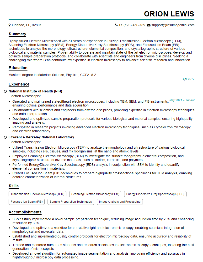
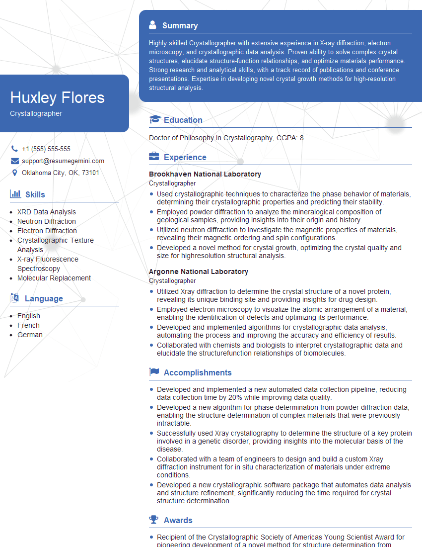
Mastering X-ray Diffraction and Electron Microscopy opens doors to exciting careers in research, development, and quality control across various industries. A strong understanding of these techniques is highly valued by employers. To significantly boost your job prospects, focus on creating a compelling and ATS-friendly resume that highlights your skills and experience effectively. ResumeGemini is a trusted resource that can help you build a professional resume tailored to your specific experience. Examples of resumes specifically tailored for candidates with expertise in X-ray Diffraction and Electron Microscopy are available through ResumeGemini to guide you in showcasing your qualifications.
Explore more articles
Users Rating of Our Blogs
Share Your Experience
We value your feedback! Please rate our content and share your thoughts (optional).
What Readers Say About Our Blog
Hi, I represent an SEO company that specialises in getting you AI citations and higher rankings on Google. I’d like to offer you a 100% free SEO audit for your website. Would you be interested?
good


