Every successful interview starts with knowing what to expect. In this blog, we’ll take you through the top Ultrasound and Imaging Techniques interview questions, breaking them down with expert tips to help you deliver impactful answers. Step into your next interview fully prepared and ready to succeed.
Questions Asked in Ultrasound and Imaging Techniques Interview
Q 1. Explain the principles of ultrasound imaging.
Ultrasound imaging, also known as sonography, relies on the principle of echolocation, similar to how bats navigate. A transducer sends high-frequency sound waves (ultrasound) into the body. These waves travel through tissues, reflecting (or echoing) back when they encounter boundaries between tissues with differing densities. The transducer receives these returning echoes, and a computer processes them to create an image. The time it takes for the sound waves to return is used to determine the depth of the structure, and the strength of the reflected signal indicates the tissue’s density. Brighter areas on the image generally represent denser tissues, while darker areas represent less dense tissues.
Think of it like shining a flashlight into a foggy room. The flashlight beam (ultrasound waves) will bounce off objects (organs and tissues) of different sizes and densities, creating shadows and highlights that reveal their shapes and positions.
Q 2. Describe the different types of ultrasound transducers and their applications.
Ultrasound transducers vary in design and frequency, each suited to specific applications. Here are a few key types:
- Linear array transducers: These produce rectangular images ideal for superficial structures like skin, muscles, and breast tissue. They offer high resolution but limited depth penetration.
- Curvilinear (convex) array transducers: These create fan-shaped images, useful for visualizing deeper structures like the abdomen, pelvis, and heart. They provide better depth penetration than linear arrays but at the cost of slightly lower resolution.
- Phased array transducers: These use electronic phasing to steer the ultrasound beam, enabling cardiac imaging from intercostal spaces or transesophageal approaches. They provide good image quality in challenging areas.
- Endocavity transducers: Small, specialized transducers designed for insertion into body cavities (rectum, vagina) providing detailed images of internal organs. They provide excellent resolution and direct visualization.
The choice of transducer depends on the specific clinical question. For example, a linear array transducer would be preferred for a thyroid ultrasound, while a curvilinear array would be better suited for a liver ultrasound.
Q 3. What are the advantages and limitations of ultrasound compared to other imaging modalities?
Ultrasound has several advantages over other imaging modalities:
- Non-invasive: It doesn’t involve ionizing radiation.
- Real-time imaging: Allows dynamic visualization of organ movement and blood flow.
- Relatively inexpensive: Compared to CT, MRI, or PET scans.
- Portable: Many ultrasound machines are portable, allowing bedside or point-of-care imaging.
However, limitations exist:
- Limited penetration: Ultrasound struggles to penetrate bone and air, limiting its use in certain areas.
- Operator-dependent: Image quality relies heavily on the skill and experience of the sonographer.
- Image quality affected by obesity: Excess adipose tissue can attenuate the ultrasound beam.
- Artifact generation: Sound wave reflections can create artifacts obscuring the underlying anatomy.
The choice of imaging modality often depends on the specific clinical scenario and the balance between advantages and limitations of each technique.
Q 4. How do you interpret ultrasound images to identify various pathologies?
Interpreting ultrasound images requires training and experience. We analyze various features:
- Echogenicity: How bright or dark a structure appears (e.g., hyperechoic – bright, hypoechoic – dark, anechoic – without echoes).
- Texture: The overall pattern of echoes (e.g., homogenous or heterogeneous).
- Shape and size: Compared to normal anatomical references.
- Borders: Well-defined or irregular borders can indicate pathology.
- Acoustic shadowing: Reduction of echoes behind highly reflective structures (e.g., stones).
- Acoustic enhancement: Increased echoes behind fluid-filled structures.
For example, a hypoechoic mass with irregular borders in the breast may suggest a malignancy, while an anechoic, well-defined structure in the kidney could represent a cyst. This interpretation is always made in clinical context, taking into account the patient’s history and physical examination findings.
Q 5. Explain the concept of acoustic impedance and its role in ultrasound imaging.
Acoustic impedance is the resistance to the propagation of sound waves in a medium. It’s determined by the density and speed of sound within the tissue. The greater the difference in acoustic impedance between two adjacent tissues, the stronger the reflection of the sound waves at their interface. This is crucial in ultrasound because it dictates the brightness of the boundaries between tissues on the image. Strong reflections generate bright echoes, representing clear tissue boundaries; minimal reflections generate weak echoes, resulting in less distinct boundaries.
For example, the strong acoustic impedance mismatch between air and soft tissue is why it’s difficult to get good ultrasound images of lung tissue. The majority of the ultrasound waves will reflect at the air-tissue interface, rather than penetrating into the lung.
Q 6. Describe the process of performing a Doppler ultrasound examination.
Doppler ultrasound uses the Doppler effect to measure the velocity of moving blood within vessels. The principle is that the frequency of the sound waves reflected from moving red blood cells changes depending on their velocity relative to the transducer. This frequency shift is analyzed by the machine to determine the blood flow velocity and direction.
The process involves positioning the transducer over a vessel, selecting the Doppler mode on the ultrasound machine (e.g., pulsed-wave or continuous-wave Doppler), and adjusting settings to optimize visualization of the blood flow. The resulting waveform displays the velocity and direction of blood flow, providing information about the patency of vessels and potential obstructions.
Pulsed-wave Doppler allows for specific depth selection, while continuous-wave Doppler provides better measurement of high-velocity flow. Color Doppler provides a color overlay on the grayscale image indicating direction and velocity of blood flow, enhancing visualization.
Q 7. How do you adjust the ultrasound machine settings to optimize image quality for different anatomical structures?
Optimizing ultrasound image quality involves adjusting several machine parameters based on the anatomical structure being imaged:
- Depth: Adjust the depth control to visualize the target structure completely, avoiding unnecessary depth and improving resolution.
- Frequency: Higher frequencies offer better resolution but less penetration, while lower frequencies provide better penetration but lower resolution. A high-frequency transducer is typically used for superficial structures, while a lower frequency transducer is used for deeper structures.
- Gain: Amplifies the received echoes, enhancing the brightness of the image. Careful adjustment is needed to avoid over-amplification which introduces noise and reduces image contrast.
- Time Gain Compensation (TGC): Compensates for the attenuation of the ultrasound beam as it travels through tissues, ensuring uniform brightness across different depths.
- Focus: Adjust the focus to optimize image clarity at a specific depth.
- Dynamic Range: Adjusts the range of gray scales displayed, influencing contrast and visibility of subtle differences in tissue echogenicity.
Each setting requires adjustment based on the specific anatomy and clinical context. A skilled sonographer can fine-tune these parameters in real-time to achieve optimal image quality, maximizing diagnostic information.
Q 8. What are the safety precautions associated with ultrasound examinations?
Ultrasound is generally considered a very safe imaging modality. However, there are some precautions to be aware of. The primary concern is the potential for bioeffects, which are biological changes caused by exposure to ultrasound waves. These are generally considered negligible at diagnostic levels, but we still adhere to the ALARA principle – As Low As Reasonably Achievable. This means we minimize exposure time and keep the output power to the minimum necessary for a diagnostic image.
- Minimizing Exposure Time: We only scan for as long as necessary to obtain the required information, avoiding unnecessary prolonging of the examination.
- Focusing the Beam: We use focused transducers to concentrate the ultrasound energy on the area of interest, reducing exposure to surrounding tissues.
- Thermal and Mechanical Bioeffects: While rare at diagnostic levels, we are mindful of the potential for these, particularly with higher intensities. This is why we avoid prolonged scanning of sensitive areas like the eyes.
- Patient Comfort: We ensure the patient is comfortable and positioned correctly to minimize movement and the need for repeated scans. This also helps maintain a good quality image and reduce the examination time.
- Operator Training and Expertise: Experienced sonographers are well-trained in ultrasound safety protocols and use their expertise to optimize image quality while minimizing exposure.
For example, during a fetal ultrasound, we would adjust the output power and scan time to minimize exposure to the developing fetus while still obtaining diagnostic images.
Q 9. Explain the artifacts commonly encountered in ultrasound imaging and their causes.
Ultrasound artifacts are errors in the image that don’t accurately represent the underlying anatomy. They can be confusing, but understanding their causes helps interpret the scan correctly. Think of them like ‘visual noise’ obscuring the true picture.
- Acoustic Shadowing: This appears as a dark area behind a highly reflective structure (e.g., a kidney stone). The sound waves are strongly reflected, preventing them from reaching deeper tissues.
- Acoustic Enhancement: The opposite of shadowing. This shows as a brighter area behind a structure that transmits sound waves exceptionally well (e.g., a fluid-filled cyst). The sound waves pass easily through the fluid, increasing the intensity of the signals behind it.
- Reverberation: This produces multiple parallel lines, often appearing as streaks or trails. It occurs when sound waves bounce back and forth between two strong reflectors, like air bubbles in the bowel.
- Mirror Image: A structure appears on the opposite side of a strong reflector. This is caused by the sound waves reflecting off the strong reflector and then being misinterpreted as coming from the other side.
- Side Lobes: These are weaker sound beams emitted from the sides of the transducer. They can create weak echoes in areas not directly in the main beam’s path, causing some blurring or misrepresentation.
For instance, recognizing acoustic shadowing behind a gallstone helps confirm its presence and differentiate it from other potential masses.
Q 10. How do you assess the quality of an ultrasound image?
Assessing ultrasound image quality is crucial for accurate diagnosis. It’s a combination of technical factors and the sonographer’s skill in optimizing the scan.
- Image Resolution: This refers to the sharpness and detail. High resolution allows us to clearly visualize small structures. We achieve this by using appropriate transducer frequencies and focusing techniques.
- Penetration: The ability to see deep structures. Factors impacting this are transducer frequency and the patient’s body habitus. Lower frequencies allow for better penetration but reduced resolution.
- Contrast Resolution: This indicates how well we can differentiate between tissues with similar acoustic impedances. Good contrast resolution is important for identifying subtle differences between normal and abnormal tissue.
- Artifacts: Their presence and type. Understanding artifacts is critical, as they can interfere with image interpretation. Minimizing artifacts improves overall image quality.
- Annotation and Labeling: Clear and accurate annotation is vital for documentation and communication between healthcare providers.
Imagine trying to read a blurry photograph versus a sharp, clear one. The same principle applies here; a high-quality ultrasound image provides significantly clearer anatomical details, leading to more accurate diagnoses.
Q 11. Describe the steps involved in preparing a patient for an ultrasound examination.
Patient preparation for an ultrasound exam varies depending on the area being scanned. The goal is to optimize image quality and ensure patient comfort.
- Abdominal Ultrasound: Patients are typically asked to fast for several hours before the exam (typically 6-8 hours) to minimize bowel gas interference. They should also void their bladder before the exam (unless a bladder scan is specifically required).
- Obstetric Ultrasound: Patients may be asked to have a full bladder to help displace the intestines and better visualize the uterus.
- Transvaginal Ultrasound: This involves a probe inserted into the vagina. It often requires no special preparation beyond a light examination.
- Cardiac Ultrasound (Echocardiography): This usually involves no specific preparation beyond ensuring comfort and possibly a resting period.
- Breast Ultrasound: Usually involves no specific preparation, though patients should wear comfortable clothing for easy access.
Clear communication with the patient about the preparation process is crucial to ensure the examination proceeds smoothly. We explain the reason for the preparation steps to increase patient compliance and reduce anxiety.
Q 12. What are the common indications for abdominal ultrasound?
Abdominal ultrasound is a versatile tool used to evaluate various organs within the abdomen. Some common indications include:
- Evaluation of abdominal pain: To identify the source of pain, such as appendicitis, cholecystitis, or kidney stones.
- Assessment of organ size and structure: To detect abnormalities in the liver, spleen, kidneys, pancreas, and gallbladder.
- Detection of masses: To identify and characterize abdominal masses, determining whether they are solid or cystic.
- Assessment of fluid collections: To detect ascites (fluid in the abdominal cavity) or abscesses.
- Evaluation of vascular structures: To visualize the aorta and other major abdominal blood vessels.
- Guidance for procedures: To guide biopsies or other minimally invasive procedures.
For example, if a patient presents with right upper quadrant pain, an abdominal ultrasound can help differentiate between gallstones, hepatitis, or other causes.
Q 13. What are the common indications for obstetric ultrasound?
Obstetric ultrasound plays a vital role in monitoring pregnancy and fetal development. Common indications include:
- Confirmation of pregnancy: Detecting the gestational sac and fetal pole early in pregnancy.
- Determination of gestational age: Measuring the fetal crown-rump length or other parameters to estimate gestational age.
- Assessment of fetal growth and development: Monitoring fetal growth, evaluating anatomy, and detecting potential abnormalities.
- Placental localization: Determining the location of the placenta to assess the risk of placenta previa or other complications.
- Assessment of amniotic fluid volume: Determining whether the fluid levels are within the normal range.
- Detection of multiple gestations: Identifying twins or higher-order multiple pregnancies.
- Assessment of fetal well-being: Monitoring fetal heart rate and movement.
Early detection of fetal anomalies through obstetric ultrasound allows for timely intervention and management, improving pregnancy outcomes.
Q 14. How do you differentiate between a solid and cystic mass on ultrasound?
Differentiating solid and cystic masses on ultrasound relies on understanding the different ways sound waves interact with tissue.
- Cystic Masses: Appear anechoic (black) because sound waves pass easily through fluid. They typically have well-defined, smooth borders and posterior acoustic enhancement (brightening behind the mass).
- Solid Masses: Appear hypoechoic (gray) or hyperechoic (bright white) depending on their composition. They usually have irregular borders and may cause acoustic shadowing (darkening behind the mass).
However, it’s crucial to remember that some masses can have mixed characteristics, making definitive identification challenging solely based on ultrasound appearance. Further investigations such as CT, MRI, or biopsy may be necessary for definitive diagnosis.
For example, a simple cyst in the ovary will typically appear anechoic with smooth borders and posterior enhancement, whereas a solid ovarian tumor might show a complex internal structure with irregular margins and potential shadowing.
Q 15. Describe the characteristic ultrasound appearance of various organs.
Ultrasound imaging relies on the reflection of sound waves to create images of internal organs. Different tissues have varying acoustic impedances, affecting how sound waves reflect. This results in distinct grayscale appearances on the ultrasound image. For example:
Liver: Typically appears homogenous (evenly textured) with a medium-grey echogenicity (brightness). Focal lesions like tumors may appear as hypoechoic (darker) or hyperechoic (brighter) areas depending on their composition.
Kidney: Shows a characteristic layered appearance. The renal cortex (outer layer) is usually isoechoic (similar brightness) to the liver, while the renal medulla (inner layer) appears hypoechoic. The renal pelvis (fluid-filled area) is anechoic (completely black, no echoes).
Spleen: Similar in echogenicity to the liver, appearing homogenous and medium-grey.
Pancreas: Often more difficult to visualize due to its location and surrounding structures. It usually appears as a relatively hypoechoic structure behind the stomach.
Gallbladder: When filled with bile, the gallbladder is anechoic. Stones within the gallbladder (choleliths) appear as hyperechoic foci with posterior acoustic shadowing (a dark area behind the stone caused by sound wave attenuation).
It’s important to remember that these are general descriptions. Variations can occur due to factors such as patient age, underlying diseases, and the specific ultrasound machine used.
Career Expert Tips:
- Ace those interviews! Prepare effectively by reviewing the Top 50 Most Common Interview Questions on ResumeGemini.
- Navigate your job search with confidence! Explore a wide range of Career Tips on ResumeGemini. Learn about common challenges and recommendations to overcome them.
- Craft the perfect resume! Master the Art of Resume Writing with ResumeGemini’s guide. Showcase your unique qualifications and achievements effectively.
- Don’t miss out on holiday savings! Build your dream resume with ResumeGemini’s ATS optimized templates.
Q 16. Explain the concept of grayscale and color Doppler imaging.
Grayscale ultrasound provides a black and white image based on the reflection of sound waves. The brightness (echogenicity) of each area on the image correlates to the acoustic impedance of the tissue. Brighter areas represent stronger reflections, and darker areas represent weaker reflections.
Color Doppler imaging adds another layer of information by showing blood flow. It uses the Doppler effect – the change in frequency of a wave due to motion – to detect movement of red blood cells. Blood flow towards the transducer is typically represented in red, while flow away is represented in blue. The color intensity reflects the velocity of blood flow.
Imagine grayscale as a photograph, showing the structure and texture. Color Doppler is like overlaying a heat map showing the direction and speed of blood flow within that structure. This combined information is crucial for evaluating vascularity and identifying abnormalities.
Q 17. What are the different types of Doppler techniques used in ultrasound?
Several Doppler techniques exist to assess blood flow:
Pulsed-wave Doppler: Uses short bursts of ultrasound waves to measure velocity at a specific point. It’s great for assessing localized flow within a vessel.
Continuous-wave Doppler: Uses continuous transmission and reception of ultrasound waves, allowing measurement of high velocities but not precise location within the vessel.
Color Doppler Imaging (CDI): Provides a two-dimensional representation of blood flow, indicating direction and velocity with color encoding. Useful for a broad overview of flow patterns.
Power Doppler Imaging (PDI): A more sensitive technique than CDI, focusing on the amplitude of the Doppler signal. It’s less sensitive to flow direction but is better at detecting low-velocity flow.
The choice of Doppler technique depends on the clinical question and the specific anatomy being assessed. For example, pulsed-wave Doppler is ideal for measuring the velocity of blood flow in a specific artery, while color Doppler provides a visual representation of flow in a larger area such as a liver.
Q 18. How do you measure blood flow velocity using Doppler ultrasound?
Doppler ultrasound measures blood flow velocity by analyzing the frequency shift (Doppler shift) of the ultrasound waves reflected from moving red blood cells. The greater the frequency shift, the higher the velocity of the blood flow. The machine uses the following principles:
Signal Acquisition: The transducer emits ultrasound waves, and the reflected waves are received. The difference between the transmitted and received frequencies is the Doppler shift.
Signal Processing: The Doppler shift is processed to calculate the blood flow velocity. This calculation involves the known frequency of the transmitted ultrasound waves and the angle of insonation (angle between the ultrasound beam and the direction of blood flow).
Velocity Display: The calculated velocity is displayed numerically or graphically on the ultrasound image. Units are usually cm/s or m/s.
Accurate velocity measurement depends on knowing the angle of insonation. A parallel angle (0 degrees) between the ultrasound beam and vessel provides the most accurate reading. Angle correction is applied if the angle is not parallel. This is a critical aspect of Doppler ultrasound technique.
Q 19. What are the potential complications associated with ultrasound examinations?
Ultrasound is generally considered a safe procedure with minimal complications. However, potential risks include:
Bioeffects: Although unlikely at diagnostic intensities, prolonged or high-intensity exposure to ultrasound could potentially cause tissue heating or cavitation (formation of bubbles in tissues). The benefits of the exam always outweigh these extremely low risks.
Infection: There is a small risk of infection at the transducer contact site, particularly if the skin is broken or compromised. Proper hygiene practices mitigate this risk.
Discomfort: Some patients may experience discomfort from pressure of the transducer on the skin, especially during prolonged examinations. This can be minimized by adjusting transducer pressure and position.
Allergic reactions: In rare cases, patients may experience allergic reactions to the gel used during the examination.
It is crucial to inform patients about the potential benefits and risks of ultrasound before the procedure.
Q 20. How do you handle challenging situations during an ultrasound exam?
Challenging situations during an ultrasound exam may include:
Suboptimal Acoustic Windows: Gas in the bowel or overlying adipose tissue can severely limit ultrasound penetration. I’d adjust transducer frequency, use different approaches, and potentially utilize harmonic imaging to overcome this.
Patient Movement: Movement artifacts can obscure the image. I’d instruct the patient to hold their breath or to remain still, and I’d employ techniques to optimize image quality despite movement.
Difficult-to-visualize Structures: Some structures are inherently challenging to image, such as the pancreas. I’d combine different scanning techniques, adjust gain and dynamic range settings, and potentially use contrast-enhanced ultrasound to improve visualization.
My approach to these challenges emphasizes patience, problem-solving, and communication with the patient to ensure the best possible examination. When faced with limitations, I clearly document the findings and any difficulties encountered in the report.
Q 21. Describe your experience with different ultrasound machines.
Throughout my career, I’ve had extensive experience operating various ultrasound machines from different manufacturers, including GE, Philips, and Siemens. Each system has its own strengths and user interface, but the underlying physics remain constant. I am proficient in operating both high-end research systems with advanced capabilities, and more basic clinical systems used for routine examinations. My experience spans across different modalities, such as general abdominal, musculoskeletal, vascular, and small parts ultrasound. I am adept at optimizing machine settings to achieve high-quality images tailored to the specific clinical situation and patient characteristics. This broad experience allows me to adapt quickly to new systems and effectively use a wide range of ultrasound applications. This ensures I can perform high-quality examinations, regardless of the available equipment.
Q 22. How do you maintain quality assurance in ultrasound imaging?
Maintaining quality assurance in ultrasound imaging is crucial for accurate diagnoses and patient safety. It involves a multi-faceted approach encompassing equipment maintenance, image optimization, and adherence to standardized protocols.
Equipment Calibration and Maintenance: Regular calibration of ultrasound machines ensures accurate measurements and consistent image quality. This includes checks on transducer performance, system settings (gain, depth, frequency), and image uniformity. We use quality control phantoms – objects with known acoustic properties – to assess the system’s performance against established standards. Any deviations require immediate attention and repair.
Image Optimization: Proper image acquisition is paramount. This involves adjusting parameters like TGC (Time Gain Compensation), focusing, and dynamic range to optimize visualization of the anatomical structures of interest. Regular training and competency assessments ensure all sonographers consistently produce high-quality images.
Standardized Protocols: Following standardized protocols for specific examinations ensures consistency and facilitates comparisons over time. This includes things like standardized image acquisition positions, depth settings, and image labeling.
Quality Control Audits: Regular internal and external quality control audits provide an objective review of our imaging practices and identify areas for improvement. This involves peer review of images and assessment of the overall quality assurance program.
For example, if a transducer shows signs of wear or inconsistent performance during phantom testing, it is immediately flagged for repair or replacement, and all images acquired with that transducer are reviewed for potential impact.
Q 23. Explain your understanding of PACS (Picture Archiving and Communication System).
PACS, or Picture Archiving and Communication System, is a centralized digital system for storing, retrieving, and distributing medical images. Think of it as a digital library specifically designed for medical images. It’s essential for efficient workflow and collaboration within healthcare settings.
Storage: PACS securely stores large volumes of medical images, including ultrasound, X-ray, CT, and MRI scans, in a readily accessible format.
Retrieval: Authorized personnel can quickly access and retrieve images from the system using various search parameters (patient name, date, study type). This eliminates the need for physical storage and facilitates efficient access to patient medical history.
Distribution: PACS enables seamless sharing of images between different departments, clinicians, and even other healthcare facilities. This facilitates efficient consultations and improved patient care.
Integration: PACS is designed to integrate with other healthcare information systems like the electronic health record (EHR), allowing for a comprehensive view of the patient’s medical information.
In practice, using PACS significantly streamlines the diagnostic process. For instance, a radiologist can review ultrasound images from different locations without having to physically transport film or wait for image transfer.
Q 24. Describe your experience with ultrasound guided procedures.
I have extensive experience with ultrasound-guided procedures. This involves using real-time ultrasound imaging to visualize anatomical structures and guide the insertion of needles or catheters during various medical procedures. It adds a level of precision that significantly improves the success rate and reduces the risk of complications.
Biopsies: Ultrasound guidance allows for precise needle placement during biopsies of organs like the liver, kidneys, or thyroid, ensuring sample acquisition from the target area.
Drainage Procedures: Ultrasound guidance helps drain fluid collections (e.g., abscesses, pleural effusions) by accurately placing a drainage catheter into the target area while minimizing the risk of damage to surrounding tissue.
Injections: Ultrasound-guided injections, such as joint injections or nerve blocks, enhance the accuracy of medication delivery, maximizing therapeutic effects and minimizing side effects.
For example, during a liver biopsy, real-time ultrasound imaging allows me to visualize the needle’s path, avoiding critical blood vessels and ensuring an optimal sample is obtained. This improves the diagnostic accuracy and minimizes the risk of complications for the patient.
Q 25. How do you maintain patient confidentiality in ultrasound imaging?
Maintaining patient confidentiality is paramount in ultrasound imaging, and I adhere strictly to all relevant regulations and protocols. This involves several key measures:
Secure Data Storage: All patient data, including images and reports, are stored in secure, password-protected servers that are compliant with HIPAA and other relevant data privacy regulations.
Access Control: Only authorized personnel with a legitimate need to access patient data can view images and reports. Access privileges are carefully managed and audited regularly.
Data Encryption: Sensitive patient data are encrypted both in transit and at rest, preventing unauthorized access even if data is intercepted.
Strict adherence to policies and procedures: This includes secure disposal of printed images and reports and following appropriate protocols for data storage and sharing.
Patient education: I ensure that patients understand how their data is being handled and what measures are in place to protect their privacy. They are always given the opportunity to ask questions and receive clarifications.
For instance, if I need to discuss a patient’s images with a colleague, I will only do so in a secure environment and always be mindful of avoiding any casual discussion of identifiable patient information in public areas.
Q 26. Explain your knowledge of relevant anatomy and physiology.
A thorough understanding of relevant anatomy and physiology is fundamental to my work as an ultrasound professional. My knowledge encompasses several key areas:
Organ Systems: I possess a detailed knowledge of the anatomy and physiology of all major organ systems, including the cardiovascular, gastrointestinal, musculoskeletal, and reproductive systems. This allows me to correctly identify and interpret ultrasound images.
Normal Anatomy: I can identify normal anatomical structures and variations in different age groups and body types. This helps me differentiate normal findings from pathological abnormalities.
Pathological Conditions: My understanding extends to the anatomical and physiological changes associated with various diseases and conditions. This enhances my ability to detect and interpret sonographic findings relevant to those pathologies.
Sonographic Anatomy: I have a strong grasp of the unique appearance of anatomical structures as visualized through ultrasound. Understanding acoustic properties of tissue (such as how sound waves are reflected and attenuated) is crucial in accurate interpretation.
For example, understanding the normal size and echogenicity of the liver is essential to detect abnormalities such as hepatomegaly or liver masses. Likewise, knowing the normal anatomy of the female pelvis is vital for accurate interpretation of gynecological ultrasound scans.
Q 27. How do you stay updated with the latest advancements in ultrasound technology?
Staying updated with the latest advancements in ultrasound technology is a continuous process that requires a multifaceted approach.
Professional Organizations: Active participation in professional organizations such as the American Institute of Ultrasound in Medicine (AIUM) provides access to continuing education opportunities, journals, and conferences that showcase the newest technologies and techniques.
Conferences and Workshops: Attending conferences and workshops allows for hands-on experience with new equipment and interactive learning from leading experts in the field.
Peer-Reviewed Journals: Regularly reviewing peer-reviewed journals and publications ensures I remain abreast of the latest research and technological developments in ultrasound imaging.
Manufacturer Training: Many ultrasound manufacturers offer training programs on their latest equipment, providing in-depth knowledge of technical specifications and optimal usage.
Online Resources: Staying updated on online resources such as medical journals, webinars, and educational platforms allows me to consistently learn and expand my knowledge.
This continuous learning ensures I can provide the best possible patient care using the most up-to-date and effective imaging techniques.
Q 28. Describe a situation where you had to troubleshoot a technical problem with an ultrasound machine.
During a routine abdominal ultrasound, the machine suddenly displayed an error message indicating a transducer malfunction. The image became distorted and unreliable.
Initial Assessment: I first checked the connections to ensure the transducer was correctly plugged into the machine. I also tried a different transducer of the same type to eliminate the possibility of a transducer-specific problem.
System Diagnostics: I then accessed the machine’s self-diagnostic menu and ran a series of tests, recording all the results. This gave me clues about the likely source of the issue.
Contacting Support: Based on the diagnostic tests, I contacted the manufacturer’s technical support team. They guided me through a series of troubleshooting steps, including verifying system software and checking internal settings.
Temporary Solution: After working through various options with the support team, we determined that a software update was required. Although the problem was resolved only after a software upgrade, the support team allowed for a temporary workaround until the update arrived, thus minimizing downtime.
Documentation: I meticulously documented the entire process, including the initial error message, diagnostic test results, troubleshooting steps, and the final resolution, adding to the database of solved problems and improving future problem-solving.
This experience highlighted the importance of systematic troubleshooting, thorough documentation, and the value of a strong technical support network in maintaining optimal equipment performance.
Key Topics to Learn for Ultrasound and Imaging Techniques Interview
- Ultrasound Physics: Understand the principles of sound wave propagation, attenuation, reflection, and scattering. Explore the different types of ultrasound transducers and their applications.
- Image Formation and Processing: Grasp the process of image formation from raw ultrasound data. Familiarize yourself with techniques like beamforming, dynamic range, and image optimization. Be prepared to discuss artifact recognition and mitigation.
- Clinical Applications of Ultrasound: Review the common uses of ultrasound in various medical specialties (e.g., abdominal, obstetrics, gynecology, cardiovascular). Focus on understanding the anatomical structures visualized and the diagnostic interpretations.
- Doppler Ultrasound: Master the principles of Doppler ultrasound, including its use in assessing blood flow velocity and direction. Understand different Doppler modes (e.g., pulsed wave, continuous wave) and their clinical applications.
- Contrast-Enhanced Ultrasound (CEUS): Learn about the principles and applications of CEUS, including the use of contrast agents to enhance image quality and improve diagnostic accuracy.
- Safety and Bioeffects: Understand the safety guidelines and potential bioeffects associated with ultrasound imaging.
- Image Interpretation and Reporting: Develop your skills in interpreting ultrasound images and preparing concise and accurate reports.
- Troubleshooting and Problem-Solving: Practice identifying and resolving common issues encountered during ultrasound examinations, such as equipment malfunctions or image artifacts.
- Emerging Technologies in Ultrasound: Stay updated on advancements in ultrasound technology, such as elastography, 3D/4D ultrasound, and fusion imaging.
- Relevant Anatomy and Physiology: Possess a strong understanding of the anatomy and physiology of the systems commonly imaged using ultrasound.
Next Steps
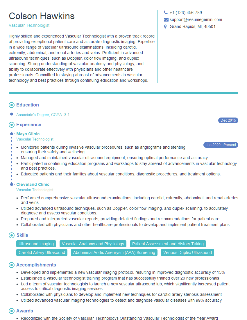
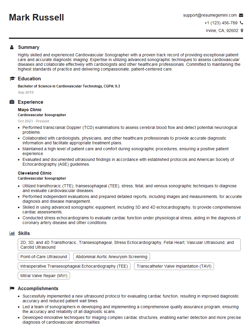
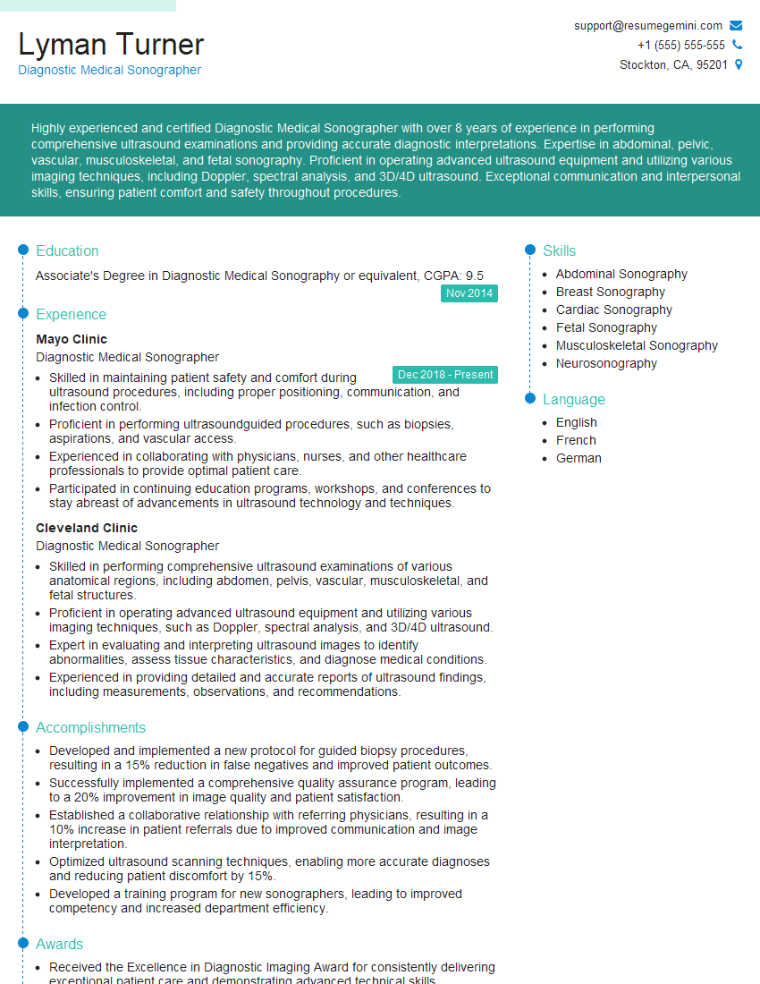
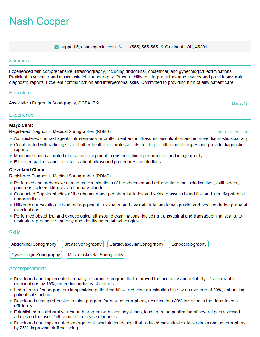
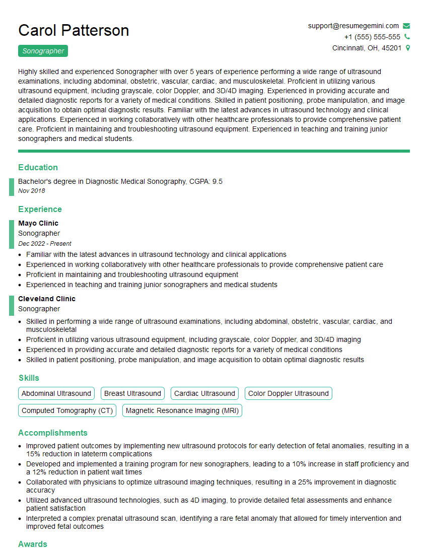
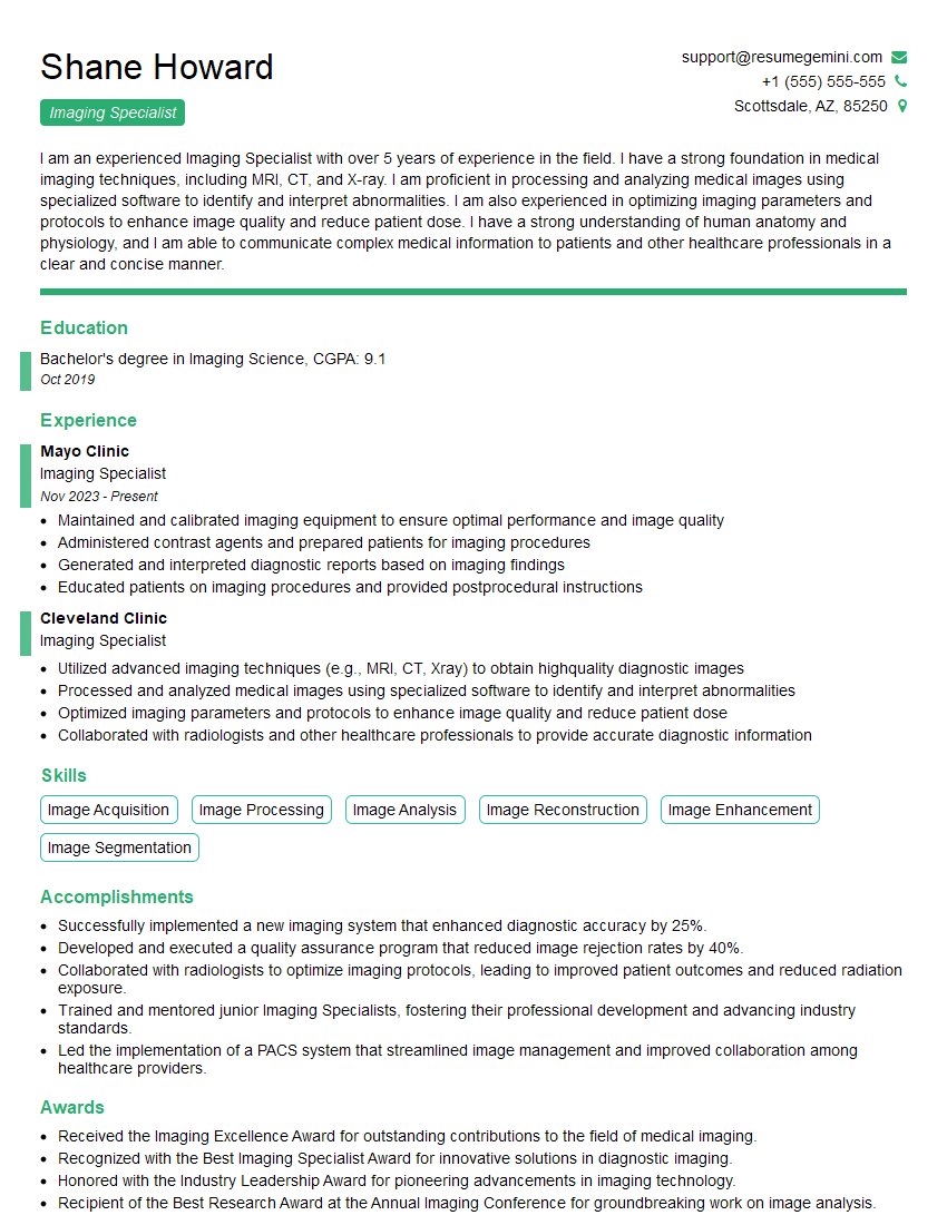
Mastering Ultrasound and Imaging Techniques is crucial for career advancement in this rapidly evolving field. A strong understanding of these concepts will significantly enhance your job prospects and open doors to exciting opportunities. To maximize your chances of landing your dream role, it’s vital to create a compelling and ATS-friendly resume that highlights your skills and experience effectively. We strongly recommend using ResumeGemini to build a professional resume that showcases your qualifications in the best possible light. ResumeGemini provides examples of resumes tailored to Ultrasound and Imaging Techniques, helping you create a document that stands out from the competition.
Explore more articles
Users Rating of Our Blogs
Share Your Experience
We value your feedback! Please rate our content and share your thoughts (optional).
What Readers Say About Our Blog
Very informative content, great job.
good